Review Article

Coronary Microvascular Spasm: Clinical Presentation and Diagnosis

Register or Login to View PDF Permissions
Permissions× For commercial reprint enquiries please contact Springer Healthcare: ReprintsWarehouse@springernature.com.

For permissions and non-commercial reprint enquiries, please visit Copyright.com to start a request.

For author reprints, please email rob.barclay@radcliffe-group.com.
Information image
Average (ratings)
No ratings
Your rating

Abstract

Professor Maseri pioneered the research and treatment of coronary vasomotion abnormalities represented by coronary vasospasm and coronary microvascular dysfunction (CMD). These mechanisms can cause myocardial ischaemia even in the absence of obstructive coronary artery disease, and have been appreciated as an important aetiology and therapeutic target with major clinical implications in patients with ischaemia with non-obstructive coronary artery disease (INOCA). Coronary microvascular spasm is one of the key mechanisms responsible for myocardial ischaemia in patients with INOCA. Comprehensive assessment of coronary vasomotor reactivity by invasive functional coronary angiography or interventional diagnostic procedure is recommended to identify the underlying mechanisms of myocardial ischaemia and to tailor the best treatment and management based on the endotype of INOCA. This review highlights the pioneering works of Professor Maseri and contemporary research on coronary vasospasm and CMD with reference to endothelial dysfunction, Rho-kinase activation and inflammation.

Disclosure:HS is on the editorial board of European Cardiology Review; this did not influence peer review. All other authors have no conflicts of interest to declare.

Received:

Accepted:

Published online:

Funding:

This work was supported, in part, by the Grant-in-Aid for Scientific Research from the Ministry of Education, Culture, Sports, Science, and Technology, Tokyo, Japan (21K16044).

Acknowledgements:We appreciate the effort of the members of the Tohoku University Hospital Cardiac Catheterization Laboratory.

Correspondence Details:Hiroaki Shimokawa, Graduate School, International University of Health and Welfare, 4-3 Kozunomori, Narita 286-8686, Japan. E: shimo@iuhw.ac.jp

Open Access:

This work is open access under the CC-BY-NC 4.0 License which allows users to copy, redistribute and make derivative works for non-commercial purposes, provided the original work is cited correctly.

Professor Maseri was a pioneer who blazed a trail in the field of coronary vasomotion abnormalities from its dawning age and was one of the most prominent physician-scientists represented in the history of the field.1,2 More than 40 years ago, he demonstrated that myocardial ischaemia can be caused by coronary vasomotion abnormalities, such as coronary vasospasm and distal coronary constriction, even in the absence of fixed obstructive coronary artery disease.3–5 Moreover, he showed that coronary vasospasm can be relieved by treatment with calcium-channel blockers, a drug of choice for contemporary vasodilator therapy in patients with vasospastic angina.6 Revascularisation of the culprit lesion has long been a cornerstone of modern care for ischaemic heart disease because of easy visibility on coronary angiography and amenability to procedural interventions by means of percutaneous coronary intervention (PCI) and coronary artery bypass grafting (CABG). However, approximately half of patients with signs and symptoms suggestive of myocardial ischaemia do not have obstructive, flow-limiting coronary artery stenosis on coronary angiography.7–16 The term ischaemia with non-obstructive coronary artery disease (INOCA) has been coined to define patients with this particular condition.17,18 The major mechanisms of myocardial ischaemia in patients with INOCA include coronary vasospasm and coronary microvascular dysfunction (CMD), the latter of which manifests itself as the structural and functional abnormalities of the coronary microvasculature, including coronary microvascular spasm.18,19 CMD is more prevalent than previously thought in many clinical settings, associated with worse clinical outcomes.12,13,20,22–33 A large-scale nationwide survey in the US evaluating a total of 12,062,081 coronary revascularisations revealed that risk-adjusted mortality did not decrease after PCI across all clinical indications.34 In line with these results, a questionable benefit of revascularisation by PCI or CABG versus optimal medical therapy in patients with stable coronary artery disease has been replicated by the findings of the two landmark clinical trials, the ORBITA trial and the ISCHEMIA trial.35,36 A recent post-hoc analysis of the ISCHEMIA trial showed that the prevalence of INOCA was at least 13% (476/3,612) in the participants with moderate or severe myocardial ischaemia, who were characterised by younger age, female preponderance, and lower atherosclerotic burden.37 Notably, the severity of ischaemia was not associated with that of non-obstructive coronary atherosclerosis.37 Although these trials did not primarily focus on coronary microvascular function, a fascinating speculation is that coronary vasomotion abnormalities, such as coronary vasospasm and CMD may contribute to myocardial ischaemia even after successful revascularisation, as predicted previously by Professor Maseri.38 This review will highlight the pioneering works of Professor Maseri and cutting-edge research on CMD from bench to bedside, with focus placed on the clinical implications of coronary microvascular spasm.

Mechanisms of Myocardial Ischaemia

The coronary circulation consists of epicardial conductive arteries (>500 μm in diameter), prearterioles (100–500 μm), arterioles (<100 μm), capillaries, and veins.39–41 Only epicardial coronary vessels are visible on coronary angiography, although most of the coronary resistance is determined by the coronary microcirculation. The mechanisms of myocardial ischaemia are three-fold: obstructive, flow-limiting stenosis of epicardial coronary arteries; epicardial coronary vasospasm; and CMD (Figure 1).19,42,43 The latter two mechanisms have been acknowledged as the major culprits of myocardial ischaemia in INOCA, although it took many years for the cardiovascular scientific community to accept the idea of vasospastic myocardial ischaemia in patients with variant angina.18,19,44–46 Armed with radioisotope techniques for measuring coronary blood flow, Professor Maseri proved that epicardial coronary vasospasm is a cause of spontaneous angina at rest in patients with variant angina.47–57 This proposal initially faced many criticisms because it contradicted the prevailing dogma that myocardial ischaemia was solely attributable to fixed obstructive coronary artery disease.46,58,59 Indeed, coronary vasospasm was once defined as “the resort of the diagnostically destitute” based on the pathological notion that atherosclerotic coronary arteries could not constrict.59 Nonetheless, an expanding body of evidence has demonstrated that coronary functional abnormalities manifested as coronary vasospasm and CMD play distinct roles in the pathophysiology of myocardial ischaemia across a wide spectrum of cardiovascular diseases.19,41,44,60–62 In the 2019 European Society of Cardiology Guidelines for the diagnosis and management of chronic coronary syndromes (CCS), patients with angina and suspected vasospastic or microvascular disease are classified as CCS V, which is one of the most frequently encountered clinical scenarios in patients with CCS.63

Mechanisms of Myocardial Ischaemia and Landmark Clinical Trials

Article image

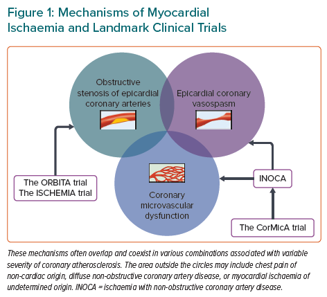
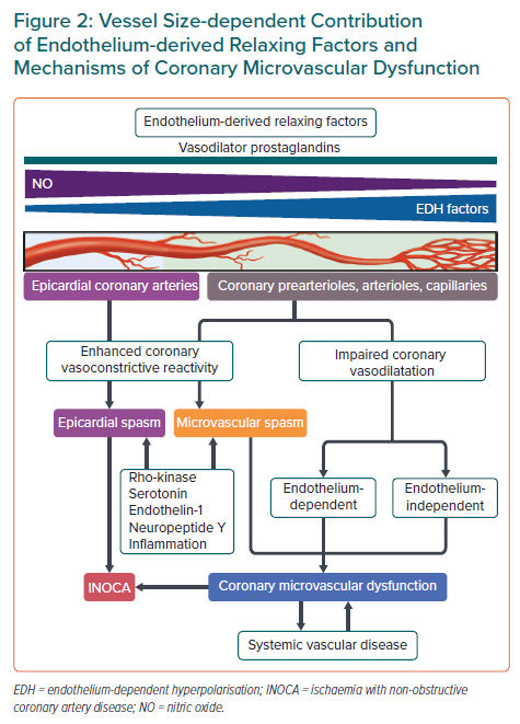
Enhanced coronary vasoconstrictive reactivity at epicardial and microvascular levels causes a transient decrease in coronary blood flow, resulting in supply ischaemia or primary angina (Figure 2).54,64 Coronary microvascular spasm is one of the mechanisms responsible for myocardial ischaemia in patients with microvascular angina (MVA).65 On the other hand, impaired endothelium-dependent and -independent coronary vasodilator capacities as well as elevated coronary microvascular resistance secondary to structural factors (e.g. luminal narrowing, vascular rarefaction, vascular remodelling and extramural compression) cause myocardial ischaemia when oxygen consumption is increased in the same way as obstructive coronary stenosis, leading to demand ischaemia or secondary angina (Figure 2).54,64 These underlying mechanisms often overlap and coexist in various combinations associated with variable degrees of coronary atherosclerosis, making the clinical picture of patients with INOCA highly heterogeneous (Figure 1).13,30,66,67 Accordingly, comprehensive assessment of coronary vasomotor reactivity by invasive functional coronary angiography or interventional diagnostic procedure using vasoactive agents (e.g. acetylcholine and adenosine) is recommended to identify the underlying mechanisms of myocardial ischaemia and to tailor the most appropriate treatment and management based on the endotype of INOCA.18,45 A good example of this tailored therapeutic strategy is provided by the CorMicA trial demonstrating that a stratified medical treatment driven by the results of coronary reactivity testing is beneficial in patients with INOCA.14 The provisional notion of Professor Maseri is certainly pertinent that a pathogenetic classification of angina may be helpful for the management of patients with the disease.54,64

Clinical Presentation of Coronary Microvascular Spasm

Coronary microvascular spasm may manifest symptomatically as MVA.65 Patients with this condition may present with typical angina, angina equivalents, such as dyspnoea and fatigue, and other atypical symptoms occurring not only at rest but also on exertion. In some cases, coronary microvascular spasm may be identified in asymptomatic subjects.65 Patients with coronary microvascular spasm are characterised by predominance of postmenopausal women, fewer coronary risk factors including smoking, and longer duration of angina.30,65,68–73 Although the prognosis of patients with MVA has been considered to be benign in most cases, the symptoms are often refractory to conventional treatment with calcium-channel blockers alone, leading to impaired quality of life.30,69,72–75 A large-scale (n=686), international, prospective cohort study by the Coronary Vasomotor Disorders International Study (COVADIS) Group recently showed that microvascular angina, in which coronary microvascular spasm was the most frequent aetiology (42%), is a considerable health problem regardless of sex or ethnicity and associated with major adverse cardiovascular events driven by hospitalisation due to unstable angina.76 The prevalence of coronary microvascular spasm ranges from approximately 2050% in patients with INOCA as well as in those with MI with non-obstructive coronary arteries, referred to as MINOCA.30,67–69,71,72,74,77–83 The mechanisms and aetiologies of INOCA are multifactorial and heterogeneous, often overlapping and coexisting in various combinations in a subclinical fashion.13,14,30,66,67,72,77 Indeed, a substantial proportion of patients with INOCA differ in the underlying aetiology of myocardial ischaemia. If complicated with CMD, the patient outcomes are at stake with increased future adverse cardiac events.20,22–33 Thus, comprehensive and multidisciplinary assessment of coronary vasomotor reactivity should be adopted to endotype patients with INOCA based on the underlying mechanisms of coronary vasomotion abnormalities for better risk stratification and management.14,18,45 Coronary microvascular spasm should not be dismissed under the umbrella of normal coronary arteries.

Diagnosis of Coronary Microvascular Spasm

The COVADIS Group has proposed a consensus set of standardised diagnostic criteria for MVA composed of four essential elements: symptoms of myocardial ischaemia; absence of obstructive coronary artery disease on coronary angiography; objective evidence of myocardial ischaemia; and evidence of impaired coronary microvascular function.65 The documentation of coronary microvascular spasm is listed as one of the standard criteria for impaired coronary microvascular function, defined as the reproduction of symptoms, ischaemic ECG changes, but no epicardial spasm during acetylcholine provocation testing.65 The diagnostic value of these criteria has been demonstrated by recent clinical studies.14,30,72,76

Vessel Size-dependent Contribution of Endothelium-derived Relaxing Factors and Mechanisms of Coronary Microvascular Dysfunction

Article image

In equivocal cases with non-diagnostic results for coronary microvascular spasm, such as reproduction of symptoms during the provocation test without ischaemic ECG changes and vice versa, myocardial ischaemia attributable to coronary microvascular spasm can be diagnosed using myocardial lactate extraction ratio or desaturation of coronary sinus blood.65,68–70,78,84,85 Lactate arises when the rate of glycolysis exceeds that of oxidation in the setting of anaerobic metabolism due to cellular hypoxia and thus transient metabolic alterations represented by myocardial lactate production serve as an objective marker of myocardial ischaemia during the spasm provocation testing.86 Briefly, blood samples are obtained simultaneously from the coronary sinus and left coronary artery during the spasm provocation testing.68,84 The net production of lactate across the coronary circulation can be calculated as (coronary sinus concentration – left coronary arterial concentration) divided by left coronary arterial concentration; a positive value indicates pathological production or release and a negative value physiological uptake or metabolism of lactate in the coronary circulation.68,84 Thrombolysis in MI (TIMI) frame count, a marker of coronary blood flow, during the spasm provocation testing may be an alternative indirect method to diagnose the occurrence of coronary microvascular spasm.78

Pathophysiology of Coronary Microvascular Dysfunction

Endothelial Modulation of Vascular Tone and Therapeutic Considerations

The endothelium, a mono-layer of endothelial cells lining the cardiovascular system, plays a pivotal role in modulating vascular tone by synthesising and liberating endothelium-derived relaxing factors (EDRFs), including vasodilator prostaglandins (e.g. prostacyclin), nitric oxide (NO), and endothelium-dependent hyperpolarisation (EDH) factors as well as endothelium-derived contracting factors (EDCFs) in response to shear stress and various agonists in vivo (Figure 2).41,44 EDH-mediated responses are observed in the presence of cyclooxygenase and NO synthase inhibitors and are accompanied with hyperpolarisation and relaxation of the underlying vascular smooth muscle cells (VSMC) and subsequent vasodilatation.41,44 Although the nature of EDH factors appears to vary depending on species and vascular beds, endothelium-derived hydrogen peroxide at physiological low concentrations plays a major role in EDH-mediated relaxations in the coronary circulation of humans and animals.87–91 In-depth information on other EDH factors is available elsewhere.41 These endothelium-derived mediators have a distinct vessel size-dependent role universally across a range of species from rodents to humans; NO predominantly mediates vasodilatation of relatively large, conduit vessels (e.g. epicardial coronary arteries), while EDH factors in small resistance vessels (e.g. coronary arterioles and microvessels) (Figure 2).41,44,92,93 Consequently, EDH-mediated relaxations are an important vasodilatory mechanism in coronary microcirculation, where coronary vascular resistance is predominantly determined. Coronary blood flow actually does not increase in response to intracoronary administration of nitroglycerin.94 EDH-mediated responses are enhanced in small resistance arteries through multiple mechanisms, including negative interactions between NO and several EDH factors.95,96

Endothelial dysfunction manifests itself as reduced production or action of EDRFs or increased responses of EDCFs.41,44 Along with coronary microvascular spasm, endothelial dysfunction contributes to the development of CMD.39–41 A large-scale cohort study (n=1,439) from the Mayo Clinic showed that as many as two-thirds of patients with INOCA had either endothelium-dependent or -independent CMD as a plausible aetiology of symptoms and signs of myocardial ischaemia, which was evaluated by invasive functional coronary angiography.13 Endothelium-dependent CMD is associated with coronary atherosclerosis and vulnerable plaque characteristics in patients with INOCA.94 Moreover, recent studies have revealed that CMD is a cardiac manifestation of the systemic small artery disease beyond the heart because of its concomitance with peripheral endothelial dysfunction.14,28,67 For example, both NO- and EDH-mediated digital vasodilatations in response to bradykinin are markedly impaired in patients with MVA.67

Contrary to a simple assumption that enhancing NO-mediated vasodilatation through supplemental NO could exert beneficial effects on patients with various cardiovascular diseases in whom the prevalence of CMD is not negligible, the effects of systemic and long-term administrations of nitrates were disappointingly unsuccessful or even harmful in patients with residual microvascular ischaemia despite successful PCI, vasospastic angina, and heart failure with preserved ejection fraction.97–100 It may be important to consider the blood vessel size-dependent contribution of NO and EDH factors in the treatment of CMD and to tailor the most appropriate therapy based on the underlying mechanisms of coronary vasomotion abnormalities (Figure 2).

Rho-kinase and Vasoconstrictive Mediators

Coronary vasoconstriction and vasospasm are the important determinants of supply ischaemia or primary angina, even in patients who have no obstructive stenosis or angiographically normal coronary arteries.44,46,101 The degree of coronary vasoconstriction is determined by the potency/efficacy of constrictor stimuli and the sensitivity/reactivity of blood vessels.46 Rho-kinase-induced myosin light chain phosphorylation with resultant VSMC hypercontraction is a major mechanism in the pathogenesis of coronary vasospasm (Figure 2).44,102–104 Intracoronary administration of fasudil, a selective Rho-kinase inhibitor, is effective for relieving refractory epicardial coronary spasm resistant to calcium-channel blockers or nitrates, coronary microvascular spasm, and the coronary slow flow phenomenon after PCI, although this drug has not yet been approved for clinical use in western countries.105–107 Moreover, Rho-kinase activity is elevated in patients with vasospastic angina who have high coronary microvascular resistance.30 Enhanced epicardial and coronary microvascular constriction/spasm is associated with increased production of vasoconstrictive mediators, including neuropeptide Y, endothelin-1, serotonin, and inflammatory responses as discussed below (Figure 2). 61,108–112 These vasoconstrictors have the potential to cause coronary artery constriction and myocardial ischaemia above the coronary autoregulation.113

Inflammation and Coronary Vasospasm

Chronic low-grade vascular inflammatory responses play important roles in the underlying mechanisms behind coronary vasospasm and CMD.61 Professor Maseri paved the way for the inflammatory hypothesis of atherosclerotic cardiovascular diseases by simply showing that patients with acute coronary syndromes who had elevated levels of C-reactive protein on admission were associated with worse outcomes.114 After a quarter century of research, this hypothesis was proved to be true by the results of the CANTOS study; treatment with canakinumab, a selective anti-interleukin-1β monoclonal antibody, significantly reduced the risk of recurrent cardiovascular events in patients with a history of MI who had a high-sensitive C-reactive protein level of 0.2 mg/dl or more as compared with placebo.115 Building on the inflammatory hypothesis, close relationships among inflammation, perivascular adipose tissue (PVAT), and vasa vasorum have emerged as key players in the pathogenesis of coronary artery spasm. A major inflammatory cytokine interleulin-1β promotes intimal thickening and coronary vasospastic responses to intracoronary serotonin or histamine via outside-to-inside signalling in pigs in vivo.116 Enhanced formation of adventitial vasa vasorum, which serves as a conduit for inflammatory mediators derived from the surrounding inflamed adipose tissue to the local coronary wall, is associated with coronary hyperconstriction via Rho-kinase activation in patients with vasospastic angina.117 Treatment with calcium-channel blockers attenuates inflammation of PVAT in the spastic coronary artery in patients with vasospastic angina in a reversible manner.118 Coronary vasospasm at epicardial and microvascular levels can be accompanied by active myocardial inflammation of cardiac sarcoidosis and immunosuppressive therapy in combination with calcium-channel blockers may be effective for alleviating the severity of coronary artery spasm in parallel with regression of myocardial inflammation of the disease.119

Conclusion

The epoch-making results of the two landmark clinical trials, the ORBITA and ISCHEMIA trials, for the management of stable coronary artery disease serve to recall the importance of coronary vasospasm and CMD beyond obstructive coronary artery disease.35,36,38,120 Professor Maseri was correct in his “search” for the origin of myocardial ischaemia in the absence of obstructive coronary arteries long before practice guidelines were published on the management of CCS including INOCA.1,2,18,45,63 He was also resourceful in his emphasis on that “elucidation of its mechanisms will lead to more appropriate therapy”.121 Cardiologists need to respond to the developing evidence irrespective of the performance measures we may adhere to in daily practice. Coronary vasomotion abnormalities will become increasingly important in the post-ISCHEMIA era. Further research is warranted to develop novel therapeutic strategies for coronary vasomotion abnormalities to improve the clinical outcomes of patients with the disease.

References

  1. Crea F, Braunwald E, Fuster V. A tribute to Attilio Maseri. Eur Heart J 2021;42:4410–2.
    Crossref
  2. Crea F, Liuzzo G. G. Attilio Maseri: scientist, clinician, mentor, and gentleman. Cardiovasc Res 2022;118:e57–8.
    Crossref | PubMed
  3. Maseri A, Pesola A, Marzilli M, et al. Coronary vasospasm in angina pectoris. Lancet 1977;309:713–7.
    Crossref | PubMed
  4. Maseri A, L’Abbate A, Baroldi G, et al. Coronary vasospasm as a possible cause of myocardial infarction. A conclusion derived from the study of “preinfarction” angina. N Engl J Med 1978;299:1271–7.
    Crossref | PubMed
  5. Pupita G, Maseri A, Kaski JC, et al. Myocardial ischemia caused by distal coronary-artery constriction in stable angina pectoris. N Engl J Med 1990;323:514–20.
    Crossref | PubMed
  6. Parodi O, Maseri A, Simonetti I. Management of unstable angina at rest by verapamil. A double-blind cross-over study in coronary care unit. Br Heart J 1979;41:167–74.
    Crossref | PubMed
  7. Bairey Merz CN, Kelsey SF, Pepine CJ, et al. The Women’s Ischemia Syndrome Evaluation (WISE) study: protocol design, methodology and feasibility report. J Am Coll Cardiol 1999;33:1453–61.
    Crossref | PubMed
  8. Shaw LJ, Bairey Merz CN, Pepine CJ, et al. Insights from the NHLBI-Sponsored Women’s Ischemia Syndrome Evaluation (WISE) study: part I: gender differences in traditional and novel risk factors, symptom evaluation, and gender-optimized diagnostic strategies. J Am Coll Cardiol 2006;47(Suppl):S4–S20.
    Crossref | PubMed
  9. Bairey Merz CN, Shaw LJ, Reis SE, et al. Insights from the NHLBI-Sponsored Women’s Ischemia Syndrome Evaluation (WISE) study: part II: gender differences in presentation, diagnosis, and outcome with regard to gender-based pathophysiology of atherosclerosis and macrovascular and microvascular coronary disease. J Am Coll Cardiol 2006;47(Suppl):S21–9.
    Crossref | PubMed
  10. Shaw LJ, Shaw RE, Bairey Merz CN, et al. Impact of ethnicity and gender differences on angiographic coronary artery disease prevalence and in-hospital mortality in the American College of Cardiology–National Cardiovascular Data Registry. Circulation 2008;117:1787–801.
    Crossref | PubMed
  11. Patel MR, Peterson ED, Dai D, et al. Low diagnostic yield of elective coronary angiography. N Engl J Med 2010;362:886–95.
    Crossref | PubMed
  12. Jespersen L, Hvelplund A, Abildstrøm SZ, et al. Stable angina pectoris with no obstructive coronary artery disease is associated with increased risks of major adverse cardiovascular events. Eur Heart J 2012;33:734–44.
    Crossref | PubMed
  13. Sara JD, Widmer RJ, Matsuzawa Y, et al. Prevalence of coronary microvascular dysfunction among patients with chest pain and nonobstructive coronary artery disease. JACC Cardiovasc Interv 2015;8:1445–53.
    Crossref | PubMed
  14. Ford TJ, Stanley B, Good R, et al. Stratified medical therapy using invasive coronary function testing in angina: the CorMicA trial. J Am Coll Cardiol 2018;72:2841–55.
    Crossref | PubMed
  15. Reeh J, Therming CB, Heitmann M, et al. Prediction of obstructive coronary artery disease and prognosis in patients with suspected stable angina. Eur Heart J 2019;40:1426–35.
    Crossref | PubMed
  16. Gitto M, Gentile F, Nowbar AN, et al. Gender-related differences in clinical presentation and angiographic findings in patients with ischemia and no obstructive coronary artery disease (INOCA): a single-center observational registry. Int J Angiol 2020;29:250–5.
    Crossref | PubMed
  17. Bairey Merz CN, Pepine CJ, Walsh MN, Fleg JL. Ischemia and no obstructive coronary artery disease (INOCA): developing evidence-based therapies and research agenda for the next decade. Circulation 2017;135:1075–92.
    Crossref | PubMed
  18. Kunadian V, Chieffo A, Camici PG, et al. An EAPCI Expert Consensus Document on ischaemia with non-obstructive coronary arteries in collaboration with European Society of Cardiology Working Group on Coronary Pathophysiology & Microcirculation Endorsed by Coronary Vasomotor Disorders International Study Group. Eur Heart J 2020;41:3504–20.
    Crossref | PubMed
  19. Shimokawa H (ed). Coronary Vasomotion Abnormalities. Singapore: Springer Singapore, 2021.
    Crossref
  20. Pepine CJ, Anderson RD, Sharaf BL, et al. Coronary microvascular reactivity to adenosine predicts adverse outcome in women evaluated for suspected ischemia results from the National Heart, Lung and Blood Institute WISE (Women’s Ischemia Syndrome Evaluation) study. J Am Coll Cardiol 2010;55:2825–32.
    Crossref | PubMed
  21. Hage C, Svedlund S, Saraste A, et al. Association of coronary microvascular dysfunction with heart failure hospitalizations and mortality in heart failure with preserved ejection fraction: a follow-up in the PROMIS-HFpEF study. J Card Fail 2020;26:1016–21.
    Crossref | PubMed
  22. von Mering GO, Arant CB, Wessel TR, et al. Abnormal coronary vasomotion as a prognostic indicator of cardiovascular events in women: results from the National Heart, Lung, and Blood Institute-Sponsored Women’s Ischemia Syndrome Evaluation (WISE). Circulation 2004;109:722–5.
    Crossref | PubMed
  23. Gulati M, Cooper-DeHoff RM, McClure C, et al. Adverse cardiovascular outcomes in women with nonobstructive coronary artery disease: a report from the Women’s Ischemia Syndrome Evaluation Study and the St James Women Take Heart Project. Arch Intern Med 2009;169:843–50.
    Crossref | PubMed
  24. Sharaf B, Wood T, Shaw L, et al. Adverse outcomes among women presenting with signs and symptoms of ischemia and no obstructive coronary artery disease: findings from the National Heart, Lung, and Blood Institute-sponsored Women’s Ischemia Syndrome Evaluation (WISE) angiographic core laboratory. Am Heart J 2013;166:134–41.
    Crossref | PubMed
  25. Maddox TM, Stanislawski MA, Grunwald GK, et al. Nonobstructive coronary artery disease and risk of myocardial infarction. JAMA 2014;312:1754–63.
    Crossref | PubMed
  26. Murthy VL, Naya M, Taqueti VR, et al. Effects of sex on coronary microvascular dysfunction and cardiac outcomes. Circulation 2014;129:2518–27.
    Crossref | PubMed
  27. Radico F, Zimarino M, Fulgenzi F, et al. Determinants of long-term clinical outcomes in patients with angina but without obstructive coronary artery disease: a systematic review and meta-analysis. Eur Heart J 2018;39:2135–46.
    Crossref | PubMed
  28. Al-Badri A, Kim JH, Liu C, et al. Peripheral microvascular function reflects coronary vascular function. Arterioscler Thromb Vasc Biol 2019;39:1492–500.
    Crossref | PubMed
  29. Lanza GA, Crea F, Kaski JC. Clinical outcomes in patients with primary stable microvascular angina: is the jury still out? Eur Heart J Qual Care Clin Outcomes 2019;5:283–91.
    Crossref | PubMed
  30. Suda A, Takahashi J, Hao K, et al. Coronary functional abnormalities in patients with angina and nonobstructive coronary artery disease. J Am Coll Cardiol 2019;74:2350–60.
    Crossref | PubMed
  31. Al-Badri A, Tahhan AS, Sabbak N, et al. Soluble urokinase-type plasminogen activator receptor and high-sensitivity troponin levels predict outcomes in nonobstructive coronary artery disease. J Am Heart Assoc 2020;9:e015515.
    Crossref | PubMed
  32. Schroder J, Michelsen MM, Mygind ND, et al. Coronary flow velocity reserve predicts adverse prognosis in women with angina and no obstructive coronary artery disease: results from the iPOWER study. Eur Heart J 2021;42:228–39.
    Crossref | PubMed
  33. Gdowski MA, Murthy VL, Doering M, et al. Association of isolated coronary microvascular dysfunction with mortality and major adverse cardiac events: a systematic review and meta-analysis of aggregate data. J Am Heart Assoc 2020;9:1-17.
    Crossref | PubMed
  34. Alkhouli M, Alqahtani F, Kalra A, et al. Trends in characteristics and outcomes of patients undergoing coronary revascularization in the United States, 2003–2016. JAMA Netw Open 2020;3:1–13.
    Crossref | PubMed
  35. Al-Lamee R, Thompson D, Dehbi HM, et al. Percutaneous coronary intervention in stable angina (ORBITA): a double-blind, randomised controlled trial. Lancet 2018;391:31–40.
    Crossref | PubMed
  36. Maron DJ, Hochman JS, Reynolds HR, et al. Initial invasive or conservative strategy for stable coronary disease. N Engl J Med 2020;382:1395–407.
    Crossref | PubMed
  37. Reynolds HR, Diaz A, Cyr DD, et al. Ischemia with nonobstructive coronary arteries: insights from the ISCHEMIA trial. JACC Cardiovasc Imaging 2022;16:63–74.
    Crossref | PubMed
  38. el-Tamimi H, Davies GJ, Sritara P, et al. Inappropriate constriction of small coronary vessels as a possible cause of a positive exercise test early after successful coronary angioplasty. Circulation 1991;84:2307–12.
    Crossref | PubMed
  39. Camici PG, Crea F. Coronary microvascular dysfunction. N Engl J Med 2007;356:830–40.
    Crossref | PubMed
  40. Camici PG, d’Amati G, Rimoldi O. Coronary microvascular dysfunction: mechanisms and functional assessment. Nat Rev Cardiol 2015;12:48–62.
    Crossref | PubMed
  41. Vanhoutte PM, Shimokawa H, Feletou M, Tang EH. Endothelial dysfunction and vascular disease - a 30th anniversary update. Acta Physiol (Oxf) 2017;219:22–96.
    Crossref | PubMed
  42. Shimokawa H, Yasuda S. Myocardial ischemia: current concepts and future perspectives. J Cardiol 2008;52:67–78.
    Crossref | PubMed
  43. Crea F, Camici PG, Bairey Merz CN. Coronary microvascular dysfunction: an update. Eur Heart J 2014;35:1101–11.
    Crossref | PubMed
  44. Shimokawa H. Williams Harvey Lecture: importance of coronary vasomotion abnormalities-from bench to bedside. Eur Heart J 2014;35:3180–93.
    Crossref | PubMed
  45. Perera D, Berry C, Hoole SP, et al. Invasive coronary physiology in patients with angina and non-obstructive coronary artery disease: a consensus document from the coronary microvascular dysfunction workstream of the British Heart Foundation/National Institute for Health Research Partnership. Heart 2022;109:88–95.
    Crossref | PubMed
  46. Maseri A, Beltrame JF, Shimokawa H. Role of coronary vasoconstriction in ischemic heart disease and search for novel therapeutic targets. Circ J 2009;73:394–403.
    Crossref | PubMed
  47. Maseri A. Pathophysiological, diagnostic and methodological problems in the study of myocardial blood flow in ischaemic heart disease. J Nucl Biol Med 1972;16:259–66.
    PubMed
  48. Maseri A, Mimmo R, Chierchia S, et al. Coronary artery spasm as a cause of acute myocardial ischemia in man. Chest 1975;68:625–33.
    Crossref | PubMed
  49. Maseri A. Pathophysiologic studies of the pulmonary and coronary circulations in man. Am J Cardiol 1976;38:751–60.
    Crossref | PubMed
  50. Maseri A, Parodi O, Severi S, Pesola A. Transient transmural reduction of myocardial blood flow demonstrated by thallium-201 scintigraphy, as a cause of variant angina. Circulation 1976;54:280–8.
    Crossref | PubMed
  51. Maseri A, Pesola A, L’Abbate A, et al. Coronary vasoconstriction as a cause of angina pectoris at rest documented by angiography and by regional myocardial perfusion radioisotopic studies. Biochem Exp Biol 1977;13:291–7.
    PubMed
  52. Maseri A, L’Abbate A, Chierchia S, Parodi O. Coronary artery spasm–diagnostic and therapeutic implications. Am Heart J 1978;96:554–5.
    Crossref | PubMed
  53. Maseri A, Severi S, Nes MD, et al. ‘Variant’ angina: one aspect of a continuous spectrum of vasospastic myocardial ischemia. Pathogenetic mechanisms, estimated incidence and clinical and coronary arteriographic findings in 138 patients. Am J Cardiol 1978;42:1019–35.
    Crossref | PubMed
  54. Maseri A, Chierchia S. Coronary vasospasm in ischemic heart disease. Chest 1980;78(Suppl):210–5.
    Crossref | PubMed
  55. Maseri A. The revival of coronary spasm. Am J Med 1981;70:752–4.
    Crossref | PubMed
  56. Deanfield JE, Maseri A, Selwyn AP, et al. Myocardial ischaemia during daily life in patients with stable angina: its relation to symptoms and heart rate changes. Lancet 1983;2:753–8.
    Crossref | PubMed
  57. Chierchia S, Gallino A, Smith G, et al. Role of heart rate in pathophysiology of chronic stable angina. Lancet 1984;2:1353–7.
    Crossref | PubMed
  58. Pickering GW. Vascular spasm. Lancet 1951;2:845–50.
    Crossref | PubMed
  59. Daley R. The autonomic nervous system in its relation to some forms of heart and lung disease. I. Heart Disease. BMJ 1957;2:173–9.
    Crossref | PubMed
  60. Del Buono MG, Montone RA, Camilli M, et al. Coronary microvascular dysfunction across the spectrum of cardiovascular diseases: JACC state-of-the-art review. J Am Coll Cardiol 2021;78:1352–71.
    Crossref | PubMed
  61. Godo S, Takahashi J, Yasuda S, Shimokawa H. Role of inflammation in coronary epicardial and microvascular dysfunction. Eur Cardiol 2021;16:e13.
    Crossref | PubMed
  62. Takahashi J, Suda A, Nishimiya K, et al. Pathophysiology and diagnosis of coronary functional abnormalities. Eur Cardiol 2021;16:e30.
    Crossref | PubMed
  63. Knuuti J, Wijns W, Saraste A, et al. 2019 ESC Guidelines for the diagnosis and management of chronic coronary syndromes. Eur Heart J 2020;41:407–77.
    Crossref | PubMed
  64. Maseri A. The changing face of angina pectoris: practical implications. Lancet 1983;1:746–9.
    Crossref | PubMed
  65. Ong P, Camici PG, Beltrame JF, et al. International standardization of diagnostic criteria for microvascular angina. Int J Cardiol 2018;250:16–20.
    Crossref | PubMed
  66. Lee BK, Lim HS, Fearon WF, et al. Invasive evaluation of patients with angina in the absence of obstructive coronary artery disease. Circulation 2015;131:1054–60.
    Crossref | PubMed
  67. Ohura-Kajitani S, Shiroto T, Godo S, et al. Marked impairment of endothelium-dependent digital vasodilatations in patients with microvascular angina: evidence for systemic small artery disease. Arterioscler Thromb Vasc Biol 2020;40:1400–12.
    Crossref | PubMed
  68. Mohri M, Koyanagi M, Egashira K, et al. Angina pectoris caused by coronary microvascular spasm. Lancet 1998;351:1165–9.
    Crossref | PubMed
  69. Masumoto A, Mohri M, Takeshita A. Three-year follow-up of the Japanese patients with microvascular angina attributable to coronary microvascular spasm. Int J Cardiol 2001;81:151–6.
    Crossref | PubMed
  70. Ohba K, Sugiyama S, Sumida H, et al. Microvascular coronary artery spasm presents distinctive clinical features with endothelial dysfunction as nonobstructive coronary artery disease. J Am Heart Assoc 2012;1:e002485.
    Crossref | PubMed
  71. Ong P, Athanasiadis A, Borgulya G, et al. Clinical usefulness, angiographic characteristics, and safety evaluation of intracoronary acetylcholine provocation testing among 921 consecutive white patients with unobstructed coronary arteries. Circulation 2014;129:1723–30.
    Crossref | PubMed
  72. Nishimiya K, Suda A, Fukui K, et al. Prognostic links between OCT-delineated coronary morphologies and coronary functional abnormalities in patients with INOCA. JACC Cardiovasc Interv 2021;14:606–18.
    Crossref | PubMed
  73. Sueda S, Sakaue T. Intracoronary ergonovine testing among 505 consecutive Japanese patients with angina-like chest pain and unobstructed coronary artery disease. Heart Vessels 2022;37:931–41.
    Crossref | PubMed
  74. Montone RA, Niccoli G, Fracassi F, et al. Patients with acute myocardial infarction and non-obstructive coronary arteries: safety and prognostic relevance of invasive coronary provocative tests. Eur Heart J 2018;39:91–8.
    Crossref | PubMed
  75. Seitz A, Feenstra R, Konst RE, et al. Acetylcholine rechallenge: a first step toward tailored treatment in patients with coronary artery spasm. JACC Cardiovasc Interv 2022;15:65–75.
    Crossref | PubMed
  76. Shimokawa H, Suda A, Takahashi J, et al. Clinical characteristics and prognosis of patients with microvascular angina: an international and prospective cohort study by the Coronary Vasomotor Disorders International Study (COVADIS) Group. Eur Heart J 2021;42:4592–600.
    Crossref | PubMed
  77. Sun H, Mohri M, Shimokawa H, et al. Coronary microvascular spasm causes myocardial ischemia in patients with vasospastic angina. J Am Coll Cardiol 2002;39:847–51.
    Crossref | PubMed
  78. Sun H, Fukumoto Y, Ito A, et al. Coronary microvascular dysfunction in patients with microvascular angina: analysis by TIMI frame count. J Cardiovasc Pharmacol 2005;46:622–6.
    Crossref | PubMed
  79. Ong P, Athanasiadis A, Borgulya G, et al. High prevalence of a pathological response to acetylcholine testing in patients with stable angina pectoris and unobstructed coronary arteries. The ACOVA study (Abnormal COronary vasomotion in patients with stable angina and unobstructed coronary arteries). J Am Coll Cardiol 2012;59:655–62.
    Crossref | PubMed
  80. Probst S, Seitz A, Martínez Pereyra V, et al. Safety assessment and results of coronary spasm provocation testing in patients with myocardial infarction with unobstructed coronary arteries compared to patients with stable angina and unobstructed coronary arteries. Eur Heart J Acute Cardiovasc Care 2020;10:380–7.
    Crossref | PubMed
  81. Mileva N, Nagumo S, Mizukami T, et al. Prevalence of coronary microvascular disease and coronary vasospasm in patients with nonobstructive coronary artery disease: systematic review and meta-analysis. J Am Heart Assoc 2022;11:e023207.
    Crossref | PubMed
  82. Aziz A, Hansen HS, Sechtem U, et al. Sex-related differences in vasomotor function in patients with angina and unobstructed coronary arteries. J Am Coll Cardiol 2017;70:2349–58.
    Crossref | PubMed
  83. Pirozzolo G, Seitz A, Athanasiadis A, et al. Microvascular spasm in non-ST-segment elevation myocardial infarction without culprit lesion (MINOCA). Clin Res Cardiol 2020;109:246–54.
    Crossref | PubMed
  84. Takahashi J, Suda A, Yasuda S, Shimokawa H. Measurement of myocardial lactate production for diagnosis of coronary microvascular spasm. J Vis Exp 2021;175:e62558.
    Crossref | PubMed
  85. Maseri A, Chierchia S. Coronary artery spasm: demonstration, definition, diagnosis, and consequences. Prog Cardiovasc Dis 1982;25:169–92.
    Crossref | PubMed
  86. Krasnow N, Neill WA, Messer JV, Gorlin R. Myocardial lactate and pyruvate metabolism. J Clin Invest 1962;41:2075–85.
    Crossref | PubMed
  87. Miura H, Bosnjak JJ, Ning G, et al. Role for hydrogen peroxide in flow-induced dilation of human coronary arterioles. Circ Res 2003;92:e31–40.
    Crossref | PubMed
  88. Matoba T, Shimokawa H, Nakashima M, et al. Hydrogen peroxide is an endothelium-derived hyperpolarizing factor in mice. J Clin Invest 2000;106:1521–30.
    Crossref | PubMed
  89. Yada T, Shimokawa H, Hiramatsu O, et al. Hydrogen peroxide, an endogenous endothelium-derived hyperpolarizing factor, plays an important role in coronary autoregulation in vivo. Circulation 2003;107:1040–5.
    Crossref | PubMed
  90. Yada T, Shimokawa H, Hiramatsu O, et al. Cardioprotective role of endogenous hydrogen peroxide during ischemia-reperfusion injury in canine coronary microcirculation in vivo. Am J Physiol Heart Circ Physiol 2006;291:H1138–46.
    Crossref | PubMed
  91. Yada T, Shimokawa H, Hiramatsu O, et al. Important role of endogenous hydrogen peroxide in pacing-induced metabolic coronary vasodilation in dogs in vivo. J Am Coll Cardiol 2007;50:1272–8.
    Crossref | PubMed
  92. Shimokawa H, Yasutake H, Fujii K, et al. The importance of the hyperpolarizing mechanism increases as the vessel size decreases in endothelium-dependent relaxations in rat mesenteric circulation. J Cardiovasc Pharmacol 1996;28:703–11.
    Crossref | PubMed
  93. Urakami-Harasawa L, Shimokawa H, Nakashima M, et al. Importance of endothelium-derived hyperpolarizing factor in human arteries. J Clin Invest 1997;100:2793–9.
    Crossref | PubMed
  94. Godo S, Corban MT, Toya T, et al. Association of coronary microvascular endothelial dysfunction with vulnerable plaque characteristics in early coronary atherosclerosis. EuroIntervention 2020;16:387–94.
    Crossref | PubMed
  95. Bauersachs J, Popp R, Hecker M, et al. Nitric oxide attenuates the release of endothelium-derived hyperpolarizing factor. Circulation 1996;94:3341–7.
    Crossref | PubMed
  96. Godo S, Sawada A, Saito H, et al. Disruption of physiological balance between nitric oxide and endothelium-dependent hyperpolarization impairs cardiovascular homeostasis in mice. Arterioscler Thromb Vasc Biol 2016;36:97–107.
    Crossref | PubMed
  97. Golino M, Spera FR, Manfredonia L, et al. Microvascular ischemia in patients with successful percutaneous coronary intervention: effects of ranolazine and isosorbide-5-mononitrate. Eur Rev Med Pharmacol Sci 2018;22:6545–50.
    Crossref | PubMed
  98. Takahashi J, Nihei T, Takagi Y, et al. Prognostic impact of chronic nitrate therapy in patients with vasospastic angina: multicentre registry study of the Japanese Coronary Spasm Association. Eur Heart J 2015;36:228–37.
    Crossref | PubMed
  99. Redfield MM, Anstrom KJ, Levine JA, et al. Isosorbide mononitrate in heart failure with preserved ejection fraction. N Engl J Med 2015;373:2314–24.
    Crossref | PubMed
  100. Borlaug BA, Anstrom KJ, Lewis GD, et al. Effect of inorganic nitrite vs placebo on exercise capacity among patients with heart failure with preserved ejection fraction: the INDIE-HFpEF randomized clinical trial. JAMA 2018;320:1764–73.
    Crossref | PubMed
  101. Shimokawa H, Tomoike H, Nabeyama S, et al. Coronary artery spasm induced in atherosclerotic miniature swine. Science 1983;221:560–2.
    Crossref | PubMed
  102. Shiroto T, Yasuda S, Tsuburaya R, et al. Role of Rho-kinase in the pathogenesis of coronary hyperconstricting responses induced by drug-eluting stents in pigs in vivo. J Am Coll Cardiol 2009;54:2321–9.
    Crossref | PubMed
  103. Kikuchi Y, Yasuda S, Aizawa K, et al. Enhanced Rho-kinase activity in circulating neutrophils of patients with vasospastic angina: a possible biomarker for diagnosis and disease activity assessment. J Am Coll Cardiol 2011;58:1231–7.
    Crossref | PubMed
  104. Shimokawa H, Sunamura S, Satoh K. RhoA/Rho-kinase in the cardiovascular system. Circ Res 2016;118:352–66.
    Crossref | PubMed
  105. Inokuchi K, Ito A, Fukumoto Y, et al. Usefulness of fasudil, a Rho-kinase inhibitor, to treat intractable severe coronary spasm after coronary artery bypass surgery. J Cardiovasc Pharmacol 2004;44:275–7.
    Crossref | PubMed
  106. Mohri M, Shimokawa H, Hirakawa Y, et al. Rho-kinase inhibition with intracoronary fasudil prevents myocardial ischemia in patients with coronary microvascular spasm. J Am Coll Cardiol 2003;41:15–9.
    Crossref | PubMed
  107. Kikuchi Y, Takahashi J, Hao K, et al. Usefulness of intracoronary administration of fasudil, a selective Rho-kinase inhibitor, for PCI-related refractory myocardial ischemia. Int J Cardiol 2019;297:8–13.
    Crossref | PubMed
  108. Clarke JG, Davies GJ, Kerwin R, et al. Coronary artery infusion of neuropeptide Y in patients with angina pectoris. Lancet 1987;1:1057–9.
    Crossref | PubMed
  109. Larkin SW, Clarke JG, Keogh BE, et al. Intracoronary endothelin induces myocardial ischemia by small vessel constriction in the dog. Am J Cardiol 1989;64:956–8.
    Crossref | PubMed
  110. Tippins JR, Antoniw JW, Maseri A. Endothelin-1 is a potent constrictor in conductive and resistive coronary arteries. J Cardiovasc Pharmacol 1989;13(Suppl 5):S177–9.
    Crossref | PubMed
  111. McFadden EP, Clarke JG, Davies GJ, et al. Effect of intracoronary serotonin on coronary vessels in patients with stable angina and patients with variant angina. N Engl J Med 1991;324:648–54.
    Crossref | PubMed
  112. Odaka Y, Takahashi J, Tsuburaya R, et al. Plasma concentration of serotonin is a novel biomarker for coronary microvascular dysfunction in patients with suspected angina and unobstructive coronary arteries. Eur Heart J 2017;38:489–96.
    Crossref | PubMed
  113. Maseri A, Crea F, Kaski JC, Crake T. Mechanisms of angina pectoris in syndrome X. J Am Coll Cardiol 1991;17:499–506.
    Crossref | PubMed
  114. Liuzzo G, Biasucci LM, Gallimore JR, et al. The prognostic value of C-reactive protein and serum amyloid A protein in severe unstable angina. N Engl J Med 1994;331:417–24.
    Crossref | PubMed
  115. Ridker PM, Everett BM, Thuren T, et al. Antiinflammatory therapy with canakinumab for atherosclerotic disease. N Engl J Med 2017;377:1119–31.
    Crossref | PubMed
  116. Shimokawa H, Ito A, Fukumoto Y, et al. Chronic treatment with interleukin-1 beta induces coronary intimal lesions and vasospastic responses in pigs in vivo. The role of platelet-derived growth factor. J Clin Invest 1996;97:769–76.
    Crossref | PubMed
  117. Nishimiya K, Matsumoto Y, Takahashi J, et al. Enhanced adventitial vasa vasorum formation in patients with vasospastic angina: assessment with OFDI. J Am Coll Cardiol 2016;67:598–600.
    Crossref | PubMed
  118. Ohyama K, Matsumoto Y, Takanami K, et al. Coronary adventitial and perivascular adipose tissue inflammation in patients with vasospastic angina. J Am Coll Cardiol 2018;71:414–25.
    Crossref | PubMed
  119. Godo S, Hasebe Y, Takahashi J, et al. Isolated cardiac sarcoidosis associated with coronary vasomotion abnormalities: a case report. Eur Heart J Case Rep 2022;6:1–7.
    Crossref | PubMed
  120. Kern MJ, Deligonul U, Vandormael M, et al. Impaired coronary vasodilator reserve in the immediate postcoronary angioplasty period: analysis of coronary artery flow velocity indexes and regional cardiac venous efflux. J Am Coll Cardiol 1989;13:860–72.
    Crossref | PubMed
  121. Maseri A, L’Abbate A, Chierchia S, et al. Significance of spasm in the pathogenesis of ischemic heart disease. Am J Cardiol 1979;44:788–92.
    Crossref | PubMed