Article

Optical Coherence Tomography For the Detection of the Vulnerable Plaque

Register or Login to View PDF Permissions
Permissions× For commercial reprint enquiries please contact Springer Healthcare: ReprintsWarehouse@springernature.com.

For permissions and non-commercial reprint enquiries, please visit Copyright.com to start a request.

For author reprints, please email rob.barclay@radcliffe-group.com.
Average (ratings)
No ratings
Your rating

Abstract

Morphological characteristics of the atheromatous plaque have been associated with the development of plaque rupture and the pathogenesis of acute coronary syndromes (ACS). Plaques with a specific morphological phenotype that are at high risk of causing ACS are called vulnerable plaques, and can be identified in vivo through the use of intracoronary imaging. Optical coherence tomography (OCT) is a high-resolution intravascular imaging modality that enables detailed visualization of atheromatous plaques. Consequently, OCT is a valuable research tool for examining the role of morphological characteristics of atheromatous plaques in the progression of coronary artery disease and plaque destabilisation, which leads to the clinical manifestation of ACS. This article summarises the pathophysiological insights obtained by OCT imaging in the formation and rupture of the vulnerable plaque.

Disclosure:AK has received research support from St. Jude Medical. KT and DT have no conflicts of interest to declare.

Received:

Accepted:

Correspondence Details:Konstantinos Toutouzas, Hippokration Hospital, 114, Vas.Sofias Avenue, 11528, Athens, Greece. E: ktoutouz@gmail.com

Copyright Statement:

The copyright in this work belongs to Radcliffe Medical Media. Only articles clearly marked with the CC BY-NC logo are published with the Creative Commons by Attribution Licence. The CC BY-NC option was not available for Radcliffe journals before 1 January 2019. Articles marked ‘Open Access’ but not marked ‘CC BY-NC’ are made freely accessible at the time of publication but are subject to standard copyright law regarding reproduction and distribution. Permission is required for reuse of this content.

Acute coronary syndromes (ACS) comprise a major cause of morbidity and mortality. The almost-exclusive cause of ACS, including unstable angina, acute myocardial infarction (AMI) with or without ST-segment elevation, and sudden cardiac death, is atherothrombosis. Pilot pathological studies have identified the pathophysiological processes implicated in the destabilisation and thrombosis of atheromatous plaques.1–3 According to the current understanding of ACS, plaques with specific morphological characteristics are more prone to rupture and development of thrombosis, which subsequently leads to the clinical manifestation of ACS. However, pathological studies are limited by their cross-sectional and teleological nature, thus imaging tools have been developed that can assess such morphological characteristics in vivo. Intracoronary imaging techniques can assess coronary plaques beyond luminal assessment (i.e. coronary angiography) and can visualise atheromatous plaques in vivo.4 Among these techniques, due to its high resolution (~15 μm) optical coherence tomography (OCT) can identify the majority of the morphological characteristics of stable and complicated atheromatous plaques, including precise measurement of the thickness of the overlying fibrous plaque and a high sensitivity for thrombus detection.5 Thus, OCT offers a valuable tool for obtaining in vivo insights into the pathomechanisms of ACS.6

Pathological Paradigm of the Vulnerable Plaque

Post-mortem studies of sudden cardiac death victims have shown that the major pathological substrates of coronary thrombosis are plaque rupture in 60–70 % of cases, plaque erosion in 20–30 % and thrombosis triggered by calcified nodules in 2–5 %.1–3 Plaque rupture, the most frequent mechanism of coronary thrombosis, involves disruption of a thin fibrous cap that overlies a large necrotic core, causing the thrombogenic contents of the necrotic core to come into contact with the bloodstream and trigger thrombus formation. Plaque erosion is defined as the presence of thrombosis in the absence of plaque thrombosis, often with absence of an endothelial layer at the thrombosed sites.2 Calcified nodules is a less common cause of coronary thrombosis, characterised by thrombus formation over nodular calcification protruding into the lumen through a disrupted thin fibrous cap.

Table 1: Sensitivity and Specificity of Optical Coherence Tomography For Detection of Different Plaque Types in Pathological Studies

Article image

Plaques that are at high risk of undergoing acute thrombosis are called vulnerable plaques and bear morphological resemblance to the aforementioned plaques.7 Nevertheless, eroded plaques do not have specific distinct morphological characteristics; therefore, the identification of erosion-prone plaques is challenging. In contrast to eroded plaques, plaques undergoing rupture have distinct morphological characteristics, including a large necrotic core, a thin (<65 μm in pathology series) and inflamed fibrous cap, high plaque burden, intimal vascularisation, inflammatory cell infiltration and spotty calcification.8 These characteristics are also observed in another type of plaque, called thin cap fibroatheroma (TCFA), which is considered the precursor of the ruptured plaque, and thus the main morphological subtype of vulnerable plaques.8

Optical Coherence Tomography Features of Stable Coronary Plaques

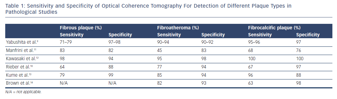
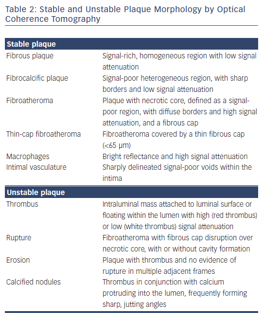
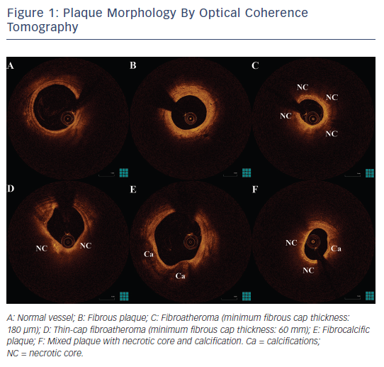
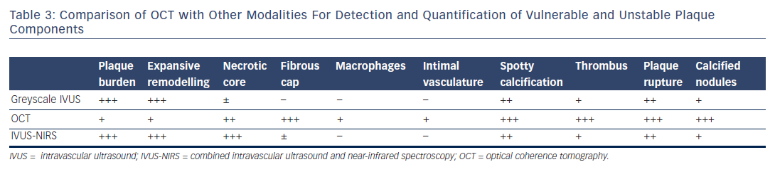
Post-mortem OCT studies have defined the optical appearance and morphological characterisation of the plaque,9–14 showing an overall good agreement with histology and high reproducibility (see Table 1). OCT can identify normal wall segments and early atherosclerotic lesions,15,16 and also perform tissue characterisation in advanced atherosclerotic lesions (see Table 2).5 The major plaque types identified by OCT are fibrous plaques, appearing as a signal-rich, homogeneous region with low signal attenuation; fibrocalcific plaques, appearing as a signal-poor heterogeneous region, with sharp borders and low signal attenuation; and necrotic core, which appears as a signal-poor region, with diffuse borders and high signal attenuation within a lesion that is covered by a fibrous cap.5,9 Fibroatheroma has been defined as a plaque with a necrotic core and a fibrous cap. Importantly, the thickness of the fibrous cap can be precisely measured by OCT.17,18 A thin-cap fibroatheroma identified by OCT is defined as a fibroatheroma covered by a thin fibrous cap; the current threshold of thin cap is the extrapolated by histology value of 65 μm, although the in vivo optimal threshold is controversial. Mixed plaques identified by OCT are those with more than one plaque morphology within a single cross-sectional frame. Additional tissue components that can be identified by OCT include macrophages that are characterised by bright reflectance and high signal attenuation, and intimal vasculature that appears as sharply delineated signal-poor voids within the intima.5,19Figure 1 demonstrates the main plaque types by OCT and a comparison of OCT with other invasive imaging modalities is presented in Table 3.

Optical Coherence Tomography Features of Unstable Coronary Plaques

Importantly, OCT can also be used for tissue characterisation in thrombosed plaques by imaging thrombus as an intraluminal mass attached to the luminal surface or floating within the lumen.20 Plaque rupture can be identified by OCT as a fibroatheroma with fibrous cap disruption over necrotic core, with or without cavity formation.5 While plaque erosion is defined as a plaque with thrombus and no evidence of rupture in multiple adjacent frames, although due to the presence of red thrombus, plaque rupture can be obscured, which can lead to an underestimation of its incidence in lesions with red thrombus. Thrombosis of calcified nodules can be identified as evidence of thrombus in conjunction with calcium protruding into the lumen, frequently forming sharp, jutting angles.21

Plaque Morphology and Clinical Presentation

As OCT can accurately image thrombosed plaques, a number of OCT studies have focused on examining pathophysiological aspects of vulnerable plaques, including mechanisms of plaque formation, progression and destabilisation. It has been shown by OCT that vulnerable plaque morphology with thin fibrous cap and OCT-detected TCFA is more frequent in unstable angina, non-ST-segment elevation myocardial infarction (NSTEMI), or ST-segment elevation myocardial infarction (STEMI) compared with stable angina.22,23 Moreover, OCT can more accurately assess complicated plaque morphology and thrombus in ACS compared with intravascular ultrasound (IVUS) or coronary angioscopy,24 enabling the evaluation of the incidence of pathomechanisms of ACS in an in vivo setting.25 In an in vivo OCT study of ACS, plaque rupture was identified in the culprit lesion of 44 % of the patients, erosion in 31 %, thrombosis of calcified nodules in 8 %, and other causes (including subocclusive stenosis, dissection and coronary spasm) in 17 %,26 showing a discordance with post-mortem studies. Of note, plaque rupture was more common in STEMI, in contrast to erosion, which was more common in patients with non-ST elevation ACS and calcified nodules that were exclusively observed in non-ST elevation ACS.

Table 2: Stable and Unstable Plaque Morphology by Optical Coherence Tomography

Article image

Figure 1: Plaque Morphology By Optical Coherence Tomography

Article image

Table 3: Comparison of OCT with Other Modalities For Detection and Quantification of Vulnerable and Unstable Plaque Components

Article image

Differences in morphological characteristics of the ruptured plaque also translate into differences in clinical presentation. Ruptured plaques of patients with STEMI are associated with a greater extent of cap disruption and a smaller minimal lumen area compared with ruptured plaques of patients with NSTEMI.27,28 Similarly, ruptured culprit plaques of ACS had a lower lumen area in comparison with non-culprit plaques in the same patients who had undergone silent rupture.29 Moreover, asymptomatic patients with silent plaque rupture on OCT examination had a higher minimal lumen area than patients with non-ST-segment elevation ACS with culprit lesion plaque rupture.30 Collectively, these studies provide indirect evidence that the pre-existing lumen narrowing might also contribute to the clinical manifestation following plaque rupture. Morphological differences in ruptured plaques have also been found between ruptured culprit plaques of patients with exercise-triggered ACS and ruptured culprit plaques of patients with rest-onset ACS, where patients with exercisetriggered ACS had a higher cap thickness and higher incidence of rupture at the shoulder of the plaque.31 Finally, the presence of culprit lesion rupture in patients with ACS has been associated with lower incidence of prodromal symptoms compared with patients without evidence of culprit plaque rupture.32

Morphology of the Vulnerable Plaque: In Vivo Measurement of Fibrous Cap

OCT has shown an association of positive remodelling of the culprit lesion with TCFA morphology and inflammation,33,34 both being important features of the vulnerable plaque as shown in pathological and in vivo IVUS studies.8,35 Furthermore, OCT has been used for identifying a critical value of cap thickness that characterises ruptureprone plaques in vivo. Although autopsy series have shown that ruptured caps have a mean thickness of 23 ± 19 μm, with 95 % being thinner than 64 μm,1 the overestimation of cap thickness by OCT in comparison with histology17 and the effect of anisotropic tissue shrinkage in histology18,36 indicate that these thresholds might not be applicable in an in vivo setting. OCT studies of ruptured plaques have shown that cap thickness in ruptured plaques can be as high as 160 μm,27,31 and it is consistently thinner in the rupture site compared with the minimal lumen area site.27 The current consensus for defining thin cap by OCT is using the histology-derived threshold of 65 μm. However, it has been shown that a fibrous cap thickness of 67 μm has only 83 % sensitivity and 77 % specificity for discriminating between ruptured and non-ruptured plaques in vivo.37 Therefore, not only fibroatheromas with cap thickness <65 μm, but also those with cap thickness ≤100 μm, could be prone to rupture.

Localisation of Vulnerable Plaques

Knowledge of spatial distribution of vulnerable plaques may lead to more effective screening of these plaques. Angiographic studies have shown that most STEMI events tend to cluster within the proximal portion of coronary artery,38 matching the distribution of coronary atherosclerosis.39 Similarly, thin-cap fibroatheromas and ruptured plaques are localised in focal spots within the proximal third of coronary art, as shown in pathological studies.40,41 OCT studies have also tried to assess plaque morphology in relation to plaque localisation. OCT can be used for assessment of plaque morphology in all coronary vessels including left main lesions,42 although imaging of true ostial lesions (i.e. ostial left main and ostial right coronary artery) might be more challenging and require more imaging attempts also with withdrawal of the guiding catheter to the aorta.43–46 Preliminary OCT studies have also demonstrated a localisation of TCFAs at the proximal segment of coronary arteries,31,47 while our group has further demonstrated that culprit lesions of patients with ACS located in the proximal 30 mm of coronary arteries were more often associated with rupture and TCFA morphology compared with the more distal lesions, indicating that plaque rupture is the main pathomechanism for ACS in these proximal lesions.48 This knowledge of the spatial distribution of the high-risk plaques can be potentially useful in the future for the development of preventive treatment strategies.49–51

Multiple Coronary Lesion Instability in Acute Coronary Syndromes

Although ACS are in principle focal manifestations arising from a single lesion, multiple lesion instability is commonly observed in atherothrombosis.52–54 Thus, non-culprit lesions of patients with MI have a more complex angiographic morphology and are associated with rapid lesion progression and increased event rates at follow-up.55,56 In addition, patients with STEMI undergoing primary percutaneous coronary intervention of the culprit lesion with additional prophylactic revascularisation of non-culprit lesion with >50 % stenosis had fewer events than patients with culprit-only treatment.57 Based on these observations, it has been hypothesised that non-culprit lesions of patients with ACS have more unstable plaque morphology than lesions of stable patients. OCT studies have further shown increased incidence of non-culprit lesion TCFA and thrombus, and a higher incidence of multiple TCFA lesions in non-culprit lesions of patients with AMI compared with non-culprit lesions of patients with stable angina.23,58 Furthermore, non-culprit lesions of patients with AMI have a higher lipid content, a thinner fibrous cap and a higher incidence of macrophage infiltration and thrombus, in conjunction with higher incidence of TCFA.59 This is mainly observed in patients with rupture of the culprit plaque as pathomechanism for the ACS,60 and is more pronounced in patients with diabetes, in whom the incidence of non-culprit lesion TCFA and rupture is higher than in those without diabetes.61

Vulnerable Plaques and Thrombolytic Therapy

In OCT studies, vulnerable plaque morphology has also been associated with impaired flow after thrombolytic therapy administration, where increased lipid content, thinner fibrous cap and plaque rupture were more common among patients without complete flow restoration compared with those with complete flow restoration. Although the reason behind this observation is not entirely clear, increased thrombogenicity of vulnerable plaques due to an exaggerated inflammatory reaction has been speculated to play an important role.62 Furthermore, in patients with STEMI with a ruptured culprit lesion, the residual thrombus burden by OCT 1 day after fibrinolysis was higher than in patients with erosion of the culprit lesion, with the presence of white thrombus at the site of the rupture,63 thus underscoring the importance of the underlying plaque morphology in STEMI as a major determinant of the response to thrombolytic therapy.

Neoatherosclerosis and the Vulnerable Plaque

The long-term follow-up of both bare metal and drug-eluting stents has demonstrated that these devices are subject to failure, and have long intervals after implantation (up to 15–20 years).64–66 Pathological studies together with in vivo OCT observations have demonstrated a variety in the neointimal patterns observed at follow-up after stent implantation, which resemble components of native atherosclerosis, and imply the atherosclerotic change of the neointimal tissue.67–71 Neoatherosclerosis has been defined by pathological studies as the presence of clusters of lipid-laden foamy macrophages with or without necrotic core formation, and/or calcification within the neointimal tissue of stented segments.68,72 Several OCT studies have demonstrated the association of neoatherosclerosis development with late stent failure.72 The role of neointimal disruption, analogue to plaque rupture in native atherosclerosis, to very late stent thrombosis is under investigation.64 Neointimal rupture has been shown to be a very prevalent mechanism in very late bare metal stent thrombosis, especially a long time after implantation,73 and is one of the most common causes of late and very late drug-eluting stent thrombosis.66,74 Neointimal rupture is a mechanism occurring mainly at longer intervals following stent implantation,75 in regions with high necrotic core content and thin fibrous cap76 (similarly to what is observed with vulnerable plaques in native atherosclerosis) and a high incidence of macrophage infiltration.76 These features are being identified mainly by OCT.72

Shortcomings of Current Vulnerable Plaque Detection Strategies: New Targets For Imaging

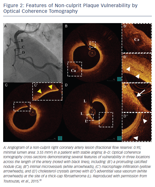
Although vulnerable plaque morphology has been prospectively associated with adverse outcomes in studies of IVUS with virtual histology and near-infrared spectroscopy, the predictive value of plaque morphology is poor, due to a high prevalence of these findings and a relatively low event rate.77–80 Consequently, despite the prognostic value of TCFA, the need to identify patients who are at high risk of cardiovascular events remains unmet.81 Therefore, new imaging targets that can enhance the prognostic potential of invasive imaging for lesion-level future events need to be identified. The identification by OCT of new imaging targets such as calcified nodules, intimal vasculature, macrophages and cholesterol crystals can offer additional information over the vulnerability of atheromatous plaques (see Figure 2). Moreover, the combination of anatomical assessment by OCT and biomechanical assessment of endothelial shear stress could give further insight into high-risk plaques.82–84

Figure 2: Features of Non-culprit Plaque Vulnerability by Optical Coherence Tomography

Article image

Figure 3: Assessment of Plaque Rupture by Polarisation-Sensitive Optical Coherence Tomography

Article image

Future Developments

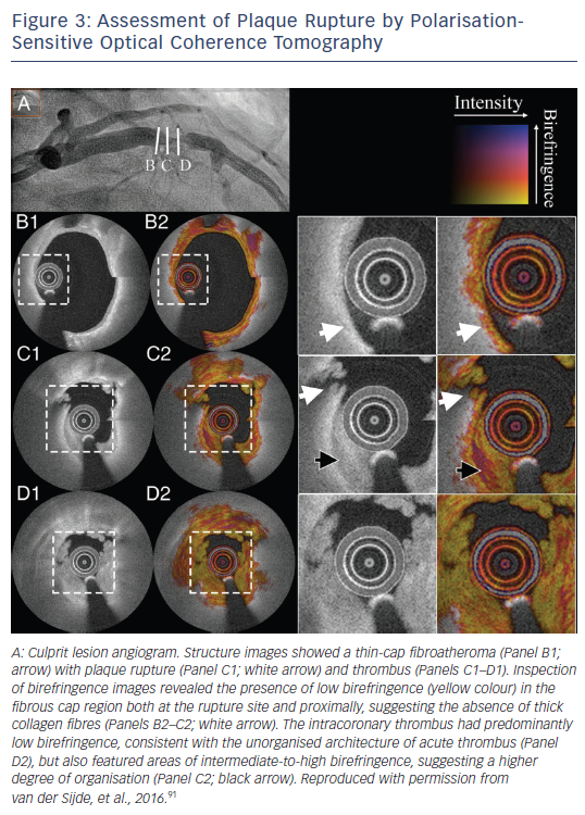
Technological advances include improved plaque characterisation using the current technology and the development of new technologies based on OCT imaging. Algorithms for the automated measurement of cap thickness are already available that can provide a fast and objective measure of this critical component of plaque vulnerability, while simultaneously increasing reproducibility of such measurements.85,86 Systems for automated tissue characterisation in atherosclerotic plaques can be used for a faster, more objective evaluation of plaque morphology.87,88 The development of ultra-high-speed OCT imaging systems will allow the complete high-resolution assessment of long arterial segments within a heartbeat,89 enabling a more accurate biomechanical assessment of the haemodynamic environment in the coronary arteries. The development of technologies such as polarisation-sensitive OCT that can measure the collagen content of atherosclerotic plaques90 will perhaps better redefine fibrous caps at a high risk of rupture (see Figure 3).91 Furthermore, new imaging systems with resolution comparable to electronic microscopy able to identify (sub-)cellular structures,92 and hybrid systems incorporating the ability for detection of molecules targeted with probes simultaneously with plaque imaging,93 are currently under development; these systems may enhance the prognostic potential of OCT imaging for vulnerable plaque identification.

Conclusion

Introduction of OCT in clinical practice has provided useful insights into the pathomechanisms of vulnerable plaque. These insights have enhanced our understanding of ACS, and have provided a knowledge base to use for clinical purposes. However, identification of the vulnerable plaque is not yet ready for clinical application, as the prognostic significance of a vulnerable plaque phenotype is poor, and a clinical benefit from interventions targeting vulnerable plaques is yet to be proven. Therefore, a better identification process needs to be implemented that can increase the prognostic significance of the findings in a way that it will be meaningful for the development and testing of local strategies targeting the vulnerable plaque. Future developments in the field will be aimed at early identification of highrisk plaques and timely intervention that could potentially result in a reduction of the mortality rates of patients with ACS.

References

  1. Burke AP, Farb A, Malcom GT, et al. Coronary risk factors and plaque morphology in men with coronary disease who died suddenly. N Engl J Med 1997;336:1276–82.
    Crossref | PubMed
  2. Falk E, Nakano M, Bentzon JF, et al. Update on acute coronary syndromes: the pathologists’ view. Eur Heart J 2013;34:719–28.
    Crossref | PubMed
  3. Virmani R, Kolodgie FD, Burke AP, et al. Lessons from sudden coronary death a comprehensive morphological classification scheme for atherosclerotic lesions. Arterioscler Thromb Vasc Biol 2000;20:1262–75.
    Crossref | PubMed
  4. Toutouzas K, Stathogiannis K, Synetos A, et al. Vulnerable atherosclerotic plaque: from the basic research laboratory to the clinic. Cardiology 2012;123:248–53.
    Crossref | PubMed
  5. Tearney GJ, Regar E, Akasaka T, et al. Consensus standards for acquisition, measurement, and reporting of intravascular optical coherence tomography studies: a report from the International Working Group for Intravascular Optical Coherence Tomography Standardization and Validation. J Am Coll Cardiol 2012;59:1058–72.
    Crossref | PubMed
  6. Karanasos A, Toutouzas K, van der Sijde J, et al. Optical coherence tomography imaging in acute myocardial infarction. In: Myocardial Infarctions. Risk Factors, Emergency Management and Long-term Health Outcomes. Nova Science Publishers, Inc. 2014; 17-52.
  7. Naghavi M, Libby P, Falk E, et al. From vulnerable plaque to vulnerable patient: a call for new definitions and risk assessment strategies: Part I. Circulation 2003;108:1664–72.
    Crossref | PubMed
  8. Virmani R, Burke AP, Farb A, Kolodgie FD. Pathology of the vulnerable plaque. J Am Coll Cardiol 2006;47:C13–8.
    Crossref | PubMed
  9. Yabushita H, Bouma BE, Houser SL, et al. Characterization of human atherosclerosis by optical coherence tomography. Circulation 2002;106:1640–5.
    Crossref | PubMed
  10. Rieber J, Meissner O, Babaryka G, et al. Diagnostic accuracy of optical coherence tomography and intravascular ultrasound for the detection and characterization of atherosclerotic plaque composition in ex-vivo coronary specimens: a comparison with histology. Coron Artery Dis 2006;17:425–30. 
    Crossref | PubMed
  11. Manfrini O, Mont E, Leone O, et al. Sources of error and interpretation of plaque morphology by optical coherence tomography. Am J Cardiol 2006;98:156–9.
    Crossref | PubMed
  12. Kawasaki M, Bouma BE, Bressner J, et al. Diagnostic accuracy of optical coherence tomography and integrated backscatter intravascular ultrasound images for tissue characterization of human coronary plaques. J Am Coll Cardiol 2006;48:81–8.
    Crossref | PubMed
  13. Kume T, Akasaka T, Kawamoto T, et al. Assessment of coronary arterial plaque by optical coherence tomography. Am J Cardiol 2006;97:1172–5.
    Crossref | PubMed
  14. Brown AJ, Obaid DR, Costopoulos C, et al. Direct comparison of virtual-histology intravascular ultrasound and optical coherence tomography imaging for identification of thin-cap fibroatheroma. Circ Cardiovasc Imaging 2015;8:e003487.
    PubMed
  15. Kume T, Akasaka T, Kawamoto T, et al. Assessment of coronary intima--media thickness by optical coherence tomography: comparison with intravascular ultrasound. Circ J 2005;69:903–7.
    Crossref | PubMed
  16. Zimarino M, Prati F, Stabile E, et al. Optical coherence tomography accurately identifies intermediate atherosclerotic lesions--an in vivo evaluation in the rabbit carotid artery. Atherosclerosis 2007;193:94–101.
    Crossref | PubMed
  17. Kume T, Akasaka T, Kawamoto T, et al. Measurement of the thickness of the fibrous cap by optical coherence tomography. Am Heart J 2006;152:755.e1–4.
    Crossref | PubMed
  18. Cilingiroglu M, Oh JH, Sugunan B, et al. Detection of vulnerable plaque in a murine model of atherosclerosis with optical coherence tomography. Catheter Cardiovasc Interv 2006;67:915–23.
    Crossref | PubMed
  19. van Soest G, Regar E, Goderie TP, et al. Pitfalls in plaque characterization by OCT: image artifacts in native coronary arteries. JACC Cardiovasc Imaging 2011;4:810–3.
    Crossref | PubMed
  20. Kume T, Akasaka T, Kawamoto T, et al. Assessment of coronary arterial thrombus by optical coherence tomography. Am J Cardiol 2006;97:1713–7.
    Crossref | PubMed
  21. Karanasos A, Ligthart JM, Witberg KT, Regar E. Calcified nodules: an underrated mechanism of coronary thrombosis? JACC Cardiovasc Imaging 2012;5:1071–2.
    Crossref | PubMed
  22. Jang I-K, Tearney GJ, MacNeill B, et al. In vivo characterization of coronary atherosclerotic plaque by use of optical coherence tomography. Circulation 2005;111:1551–5.
    Crossref | PubMed
  23. Fujii K, Masutani M, Okumura T, et al. Frequency and predictor of coronary thin-cap fibroatheroma in patients with acute myocardial infarction and stable angina pectoris a 3-vessel optical coherence tomography study. J Am Coll Cardiol 2008;52:787–8.
    Crossref | PubMed
  24. Kubo T, Imanishi T, Takarada S, et al. Assessment of culprit lesion morphology in acute myocardial infarction: ability of optical coherence tomography compared with intravascular ultrasound and coronary angioscopy. J Am Coll Cardiol 2007;50:933–9.
    Crossref | PubMed
  25. Gonzalo N, Tearney GJ, van Soest G, et al. Witnessed coronary plaque rupture during cardiac catheterization. JACC Cardiovasc Imaging 2011;4:437–8.
    Crossref | PubMed
  26. Jia H, Abtahian F, Aguirre AD, et al. In vivo diagnosis of plaque erosion and calcified nodule in patients with acute coronary syndrome by intravascular optical coherence tomography. J Am Coll Cardiol 2013;62:1748–58. 
    Crossref | PubMed
  27. Toutouzas K, Karanasos A, Tsiamis E, et al. New insights by optical coherence tomography into the differences and similarities of culprit ruptured plaque morphology in non-STelevation myocardial infarction and ST-elevation myocardial infarction. Am Heart J 2011;161:1192–9.
    Crossref | PubMed
  28. Ino Y, Kubo T, Tanaka A, et al. Difference of culprit lesion morphologies between ST-segment elevation myocardial infarction and non-ST-segment elevation acute coronary syndrome: an optical coherence tomography study. JACC Cardiovasc Interv 2011;4:76–82. 
    Crossref | PubMed
  29. Tian J, Ren X, Vergallo R, et al. Distinct morphological features of ruptured culprit plaque for acute coronary events compared to those with silent rupture and thin-cap fibroatheroma: a combined optical coherence tomography and intravascular ultrasound study. J Am Coll Cardiol 2014;63:2209–16. 
    Crossref | PubMed
  30. Shimamura K, Ino Y, Kubo T, et al. Difference of ruptured plaque morphology between asymptomatic coronary artery disease and non-st elevation acute coronary syndrome patients: an optical coherence tomography study. Atherosclerosis 2014;235:532–7. 
    Crossref | PubMed
  31. Tanaka A, Imanishi T, Kitabata H, et al. Morphology of exertion-triggered plaque rupture in patients with acute coronary syndrome: an optical coherence tomography study. Circulation 2008;118:2368–73. 
    Crossref | PubMed
  32. Kato M, Dote K, Sasaki S, et al. Presentations of acute coronary syndrome related to coronary lesion morphologies as assessed by intravascular ultrasound and optical coherence tomography. Int J Cardiol 2013;165:506–11.
    Crossref | PubMed
  33. Raffel OC, Merchant FM, Tearney GJ, et al. In vivo association between positive coronary artery remodelling and coronary plaque characteristics assessed by intravascular optical coherence tomography. Eur Heart J 2008;29:1721–8. 
    Crossref | PubMed
  34. Kashiwagi M, Tanaka A, Kitabata H, et al. Relationship between coronary arterial remodeling, fibrous cap thickness and high-sensitivity C-reactive protein levels in patients with acute coronary syndrome. Circ J 2009;73:1291–5.
    Crossref | PubMed
  35. Toutouzas K, Synetos A, Stefanadi E, et al. Correlation between morphologic characteristics and local temperature differences in culprit lesions of patients with symptomatic coronary artery disease. J Am Coll Cardiol 2007;49:2264–71.
    Crossref | PubMed
  36. Barlis P, Serruys PW, Gonzalo N, et al. Assessment of culprit and remote coronary narrowings using optical coherence tomography with long-term outcomes. Am J Cardiol 2008;102:391–5.
    Crossref | PubMed
  37. Yonetsu T, Kakuta T, Lee T, et al. In vivo critical fibrous cap thickness for rupture-prone coronary plaques assessed by optical coherence tomography. Eur Heart J 2011;32:1251–9.
    Crossref | PubMed
  38. Wang JC, Normand S-LT, Mauri L, Kuntz RE. Coronary artery spatial distribution of acute myocardial infarction occlusions. Circulation 2004;110:278–84.
    Crossref | PubMed
  39. Hochman JS, Phillips WJ, Ruggieri D, Ryan SF. The distribution of atherosclerotic lesions in the coronary arterial tree: relation to cardiac risk factors. Am Heart J 1988;116:1217–22. 
    Crossref | PubMed
  40. Cheruvu PK, Finn AV, Gardner C, et al. Frequency and distribution of thin-cap fibroatheroma and ruptured plaques in human coronary arteries: a pathologic study. J Am Coll Cardiol 2007;50:940–9.
    Crossref | PubMed
  41. Kume T, Okura H, Yamada R, et al. Frequency and spatial distribution of thin-cap fibroatheroma assessed by 3-vessel intravascular ultrasound and optical coherence tomography: an ex vivo validation and an initial in vivo feasibility study. Circ J 2009;73:1086–91.
    Crossref | PubMed
  42. van der Sijde JN, Karanasos A, van Ditzhuijzen NS, et al. Safety of optical coherence tomography in daily practice: a comparison with intravascular ultrasound. Eur Heart J Cardiovasc Imaging 2016. pii: jew037. [Epub ahead of print].
    Crossref | PubMed
  43. Fujino Y, Bezerra HG, Attizzani GF, et al. Frequencydomain optical coherence tomography assessment of unprotected left main coronary artery disease-a comparison with intravascular ultrasound. Catheter Cardiovasc Interv 2013;82:E173–83.
    Crossref | PubMed
  44. Burzotta F, Dato I, Trani C, et al. Frequency domain optical coherence tomography to assess non-ostial left main coronary artery. EuroIntervention 2015;10:e1–8.
    Crossref | PubMed
  45. Dato I, Burzotta F, Trani C, et al. Angiographically intermediate left main bifurcation disease assessment by frequency domain optical coherence tomography (FD-OCT). Int J Cardiol 2016;220:726–8.
    Crossref | PubMed
  46. Parodi G, Maehara A, Giuliani G, et al. Optical coherence tomography in unprotected left main coronary artery stenting. EuroIntervention 2010;6:94–9.
    Crossref | PubMed
  47. Fujii K, Kawasaki D, Masutani M, et al. OCT assessment of thin-cap fibroatheroma distribution in native coronary arteries. JACC Cardiovasc Imaging 2010;3:168–75.
    Crossref | PubMed
  48. Toutouzas K, Karanasos A, Riga M, et al. Optical coherence tomography assessment of the spatial distribution of culprit ruptured plaques and thin-cap fibroatheromas in acute coronary syndrome. EuroIntervention 2012;8:477–85.
    Crossref | PubMed
  49. Karanasos A, Simsek C, Serruys P, et al. Five-year optical coherence tomography follow-up of an everolimus-eluting bioresorbable vascular scaffold: changing the paradigm of coronary stenting? Circulation 2012;126:e89–91.
    Crossref | PubMed
  50. Wykrzykowska JJ, Diletti R, Gutierrez-Chico JL, et al. Plaque sealing and passivation with a mechanical self-expanding low outward force nitinol vShield device for the treatment of IVUS and OCT-derived thin cap fibroatheromas (TCFAs) in native coronary arteries: report of the pilot study vShield Evaluated at Cardiac hospital in Rotterdam for Investigation and Treatment of TCFA (SECRITT). EuroIntervention 2012;8:945–54.
    Crossref | PubMed
  51. Karanasos A, Simsek C, Gnanadesigan M, et al. OCT assessment of the long-term vascular healing response 5 years after everolimus-eluting bioresorbable vascular scaffold. J Am Coll Cardiol 2014;64:2343–56.
    Crossref | PubMed
  52. Toutouzas K, Drakopoulou M, Markou V, et al. Correlation of systemic inflammation with local inflammatory activity in non-culprit lesions: beneficial effect of statins. Int J Cardiol 2007;119:368–73.
    Crossref | PubMed
  53. Toutouzas K, Drakopoulou M, Mitropoulos J, et al. Elevated plaque temperature in non-culprit de novo atheromatous lesions of patients with acute coronary syndromes. J Am Coll Cardiol 2006;47:301–6.
    Crossref | PubMed
  54. Toutouzas K, Drakopoulou M, Vaina S, Stefanadis C. Significance of characterization of non-culprit lesions: an underscored clinical problem. Atherosclerosis 2007;195: 236–41.
    Crossref | PubMed
  55. Goldstein JA, Demetriou D, Grines CL, et al. Multiple complex coronary plaques in patients with acute myocardial infarction. N Engl J Med 2000;343:915–22.
    Crossref | PubMed
  56. Tsiamis E, Toutouzas K, Synetos A, et al. Prognostic clinical and angiographic characteristics for the development of a new significant lesion in remote segments after successful percutaneous coronary intervention. Int J Cardiol 2010;143: 29–34.
    Crossref | PubMed
  57. Wald DS, Morris JK, Wald NJ, et al. Randomized trial of preventive angioplasty in myocardial infarction. N Engl J Med 2013;369:1115–23.
    Crossref | PubMed
  58. Kubo T, Imanishi T, Kashiwagi M, et al. Multiple coronary lesion instability in patients with acute myocardial infarction as determined by optical coherence tomography. Am J Cardiol 2010;105:318–22.
    Crossref | PubMed
  59. Kato K, Yonetsu T, Kim S-J, et al. Nonculprit plaques in patients with acute coronary syndromes have more vulnerable features compared with those with non-acute coronary syndromes: a 3-vessel optical coherence tomography study. Circ Cardiovasc Imaging 2012;5:433–40. 
    Crossref | PubMed
  60. Vergallo R, Ren X, Yonetsu T, et al. Pancoronary plaque vulnerability in patients with acute coronary syndrome and ruptured culprit plaque: a 3-vessel optical coherence tomography study. Am Heart J 2014;167:59–67.
    Crossref | PubMed
  61. Fukunaga M, Fujii K, Nakata T, et al. Multiple complex coronary atherosclerosis in diabetic patients with acute myocardial infarction: a three-vessel optical coherence tomography study. EuroIntervention 2012;8:955–61.
    Crossref | PubMed
  62. Toutouzas K, Tsiamis E, Karanasos A, et al. Morphological characteristics of culprit atheromatic plaque are associated with coronary flow after thrombolytic therapy: new implications of optical coherence tomography from a multicenter study. JACC Cardiovasc Interv 2010;3:507–14.
    Crossref | PubMed
  63. Hu S, Yonetsu T, Jia H, et al. Residual thrombus pattern in patients with ST-segment elevation myocardial infarction caused by plaque erosion versus plaque rupture after successful fibrinolysis: an optical coherence tomography study. J Am Coll Cardiol 2014;63:1336–8. 
    Crossref | PubMed
  64. Karanasos A, Ligthart JM, Regar E. In-stent neoatherosclerosis: a cause of late stent thrombosis in a patient with “full metal jacket” 15 years after implantation: insights from optical coherence tomography. JACC Cardiovasc Interv 2012;5:799–800.
    Crossref | PubMed
  65. Yamaji K, Kimura T, Morimoto T, et al. Very long-term (15 to 20 years) clinical and angiographic outcome after coronary bare metal stent implantation. Circ Cardiovasc Interv 2010;3:468–75. 
    Crossref | PubMed
  66. Taniwaki M, Radu MD, Zaugg S, et al. Mechanisms of very late drug-eluting stent thrombosis assessed by optical coherence tomography. Circulation 2016;133:650–60.
    Crossref | PubMed
  67. Inoue K, Abe K, Ando K, et al. Pathological analyses of longterm intracoronary Palmaz-Schatz stenting; Is its efficacy permanent? Cardiovasc Pathol 2004;13:109–15.
    Crossref | PubMed
  68. Nakazawa G, Otsuka F, Nakano M, et al. The pathology of neoatherosclerosis in human coronary implants bare-metal and drug-eluting stents. J Am Coll Cardiol 2011;57:1314–22.
    Crossref | PubMed
  69. Gonzalo N, Serruys PW, Okamura T, et al. Optical coherence tomography patterns of stent restenosis. Am Heart J 2009;158:284–93.
    Crossref | PubMed
  70. Hou J, Qi H, Zhang M, et al. Development of lipid-rich plaque inside bare metal stent: possible mechanism of late stent thrombosis? An optical coherence tomography study. Heart 2010;96:1187–90.
    Crossref | PubMed
  71. Tanimoto S, Aoki J, Serruys PW, Regar E. Paclitaxel-eluting stent restenosis shows three-layer appearance by optical coherence tomography. EuroIntervention 2006;1:484.
    PubMed
  72. Zhang BC, Karanasos A, Regar E. OCT demonstrating neoatherosclerosis as part of the continuous process of coronary artery disease. Herz 2015;40:845–54.
    Crossref | PubMed
  73. Karanasos A, Witberg K, Ligthart J, et al. In-stent neoatherosclerosis: Are first generation drug eluting stents different than bare metal stents? An optical coherence tomography study. Circulation 2012;126:A19296.
  74. Souteyrand G, Amabile N, Mangin L, et al. Mechanisms of stent thrombosis analysed by optical coherence tomography: insights from the national PESTO French registry. Eur Heart J 2016;37:1208–16.
    Crossref | PubMed
  75. Amabile N, Souteyrand G, Ghostine S, et al. Very late stent thrombosis related to incomplete neointimal coverage or neoatherosclerotic plaque rupture identified by optical coherence tomography imaging. Eur Heart J Cardiovasc Imaging 2014;15:24–31.
    Crossref | PubMed
  76. Karanasos A, Ligthart J, Witberg K, et al. Association of neointimal morphology by optical coherence tomography with rupture of neoatherosclerotic plaque very late after coronary stent implantation. Proc. SPIE 8565, Photonic Therapeutics and Diagnostics IX, 856542 2013.
    Crossref
  77. Calvert PA, Obaid DR, O’Sullivan M, et al. Association between IVUS findings and adverse outcomes in patients with coronary artery disease: the VIVA (VH-IVUS in Vulnerable Atherosclerosis) Study. JACC Cardiovasc Imaging 2011;4: 894–901.
    Crossref | PubMed
  78. Oemrawsingh RM, Cheng JM, Garcia-Garcia HM, et al. Nearinfrared spectroscopy predicts cardiovascular outcome in patients with coronary artery disease. J Am Coll Cardiol 2014;64:2510–8.
    Crossref | PubMed
  79. Stone GW, Maehara A, Lansky AJ, et al. A prospective naturalhistory study of coronary atherosclerosis. N Engl J Med 2011;364:226–35.
    Crossref | PubMed
  80. Cheng JM, Garcia-Garcia HM, de Boer SP, et al. In vivo detection of high-risk coronary plaques by radiofrequency intravascular ultrasound and cardiovascular outcome: results of the ATHEROREMO-IVUS study. Eur Heart J 2014;35:639–47.
    Crossref | PubMed
  81. Toutouzas K, Benetos G, Karanasos A, et al. Vulnerable plaque imaging: updates on new pathobiological mechanisms. Eur Heart J 2015;36:3147–54.
    Crossref | PubMed
  82. Chatzizisis YS, Toutouzas K, Giannopoulos AA, et al. Association of global and local low endothelial shear stress with high-risk plaque using intracoronary 3D optical coherence tomography: Introduction of ‘shear stress score’. Eur Heart J Cardiovasc Imaging 2016; pii: jew134. [Epub ahead of print]. 
    Crossref | PubMed
  83. Toutouzas K, Chatzizisis YS, Riga M, et al. Accurate and reproducible reconstruction of coronary arteries and endothelial shear stress calculation using 3D OCT: comparative study to 3D IVUS and 3D QCA. Atherosclerosis 2015;240:510–9.
    Crossref | PubMed
  84. Zahnd G, Schrauwen J, Karanasos A, et al. Fusion of fibrous cap thickness and wall shear stress to assess plaque vulnerability in coronary arteries: a pilot study. Int J Comput Assist Radiol Surg 2016;11:1779–90.
    Crossref | PubMed
  85. Wang Z, Chamie D, Bezerra HG, et al. Volumetric quantification of fibrous caps using intravascular optical coherence tomography. Biomed Opt Express 2012;3:1413–26.
    Crossref | PubMed
  86. Zahnd G, Karanasos A, van Soest G, et al. Quantification of fibrous cap thickness in intracoronary optical coherence tomography with a contour segmentation method based on dynamic programming. Int J Comput Assist Radiol Surg 2015;10:1383–94.
    Crossref | PubMed
  87. Ughi GJ, Adriaenssens T, Sinnaeve P, et al. Automated tissue characterization of in vivo atherosclerotic plaques by intravascular optical coherence tomography images. Biomed Opt Express 2013;4:1014–30.
    Crossref | PubMed
  88. Gnanadesigan M, Hussain AS, White S, et al. Optical coherence tomography attenuation imaging for lipid core detection: an ex-vivo validation study. Int J Cardiovasc Imaging 2016. [Epub ahead of print].
    Crossref | PubMed
  89. Wang T, Wieser W, Springeling G, et al. Intravascular optical coherence tomography imaging at 3200 frames per second. Opt Lett 2013;38:1715–7.
    Crossref | PubMed
  90. Nadkarni SK, Pierce MC, Park BH, et al. Measurement of collagen and smooth muscle cell content in atherosclerotic plaques using polarization-sensitive optical coherence tomography. J Am Coll Cardiol 2007;49:1474–81.
    Crossref | PubMed
  91. van der Sijde JN, Karanasos A, Villiger M, et al. First-in-man assessment of plaque rupture by polarization-sensitive optical frequency domain imaging in vivo. Eur Heart J 2016;37:1932.
    Crossref | PubMed
  92. Liu L, Gardecki JA, Nadkarni SK, et al. Imaging the subcellular structure of human coronary atherosclerosis using microoptical coherence tomography. Nat Med. 2011;17:1010-4.
    Crossref | PubMed
  93. Yoo H, Kim JW, Shishkov M, et al. Intra-arterial catheter for simultaneous microstructural and molecular imaging in vivo. Nat Med 2011;17:1680–4.
    Crossref | PubMed