Article

"Save the Last Dance" for Cardiovascular Magnetic Resonance

Register or Login to View PDF Permissions
Permissions× For commercial reprint enquiries please contact Springer Healthcare: ReprintsWarehouse@springernature.com.

For permissions and non-commercial reprint enquiries, please visit Copyright.com to start a request.

For author reprints, please email rob.barclay@radcliffe-group.com.
Average (ratings)
No ratings
Your rating

Abstract

Despite high mortality, cardiovascular disease (CVD) is underestimated in autoimmune rheumatic diseases (ARDs), due to its atypical presentation. The multi-faceted nature of CVD in ARDs created the need of a dedicated outpatient cardio-rheumatic clinic. Clinical examination, rest/exercise ECG, echocardiography, nuclear techniques and cardiac catheterisation were used as first-line diagnostic tools. Although the currently used non-invasive modalities perform well in cardiology, they are unable to diagnose the complex CVD pathophysiology of ARDs. The application of cardiovascular magnetic resonance (CMR) offers some significant advantages. CMR is versatile and can be used to perform functional, stress-rest perfusion, fibrosis and evaluation of great, peripheral and coronary vessels patency, without the use of ionising radiation, allowing early diagnosis of CVD and prompting modifications of anti-rheumatic and cardiac treatment.

Disclosure:The authors have no conflicts of interest to declare.

Received:

Accepted:

Correspondence Details:Sophie Mavrogeni, 50 Esperou Street, 175–61 P. Faliro, Athens, Greece. E: soma13@otenet.gr

Copyright Statement:

The copyright in this work belongs to Radcliffe Medical Media. Only articles clearly marked with the CC BY-NC logo are published with the Creative Commons by Attribution Licence. The CC BY-NC option was not available for Radcliffe journals before 1 January 2019. Articles marked ‘Open Access’ but not marked ‘CC BY-NC’ are made freely accessible at the time of publication but are subject to standard copyright law regarding reproduction and distribution. Permission is required for reuse of this content.

Rheumatoid arthritis, the spondyloarthropathies, systemic lupus erythematosus, systemic vasculitides, inflammatory myopathies, systemic sclerosis and mixed connective tissue disease are autoimmune rheumatic diseases (ARDs) with high incidence of cardiovascular disease (CVD).1 CVD is usually underestimated in patients with rheumatic diseases, because the main focus of rheumatologists is the signs and symptoms of the systemic disease. Although targeted treatment has significantly contributed to the decrease in disease-related mortality, life expectancy in people with ARDs remains lower compared to the general population, mainly due to increased cardiovascular involvement.2–7

CVD in ARDs is the result of various pathophysiologic mechanisms including systemic, myocardial, vascular inflammation, macro- and micro-vascular ischemia, abnormal coronary vaso-reactivity and diffuse or replacement myocardial fibrosis.8,9 Irrespective of pathophysiologic background, the symptoms of heart involvement in ARDs are subtle and usually underestimated because they are attributed to the underlying systemic disease. However, the development of clinically overt cardiac signs indicates advanced cardiac disease and carries a poor prognosis.10

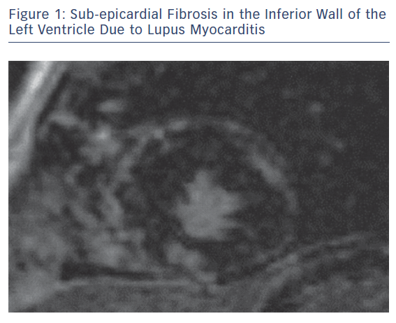
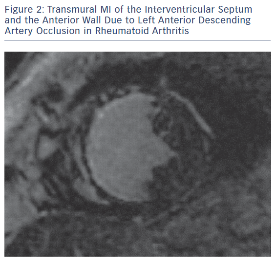
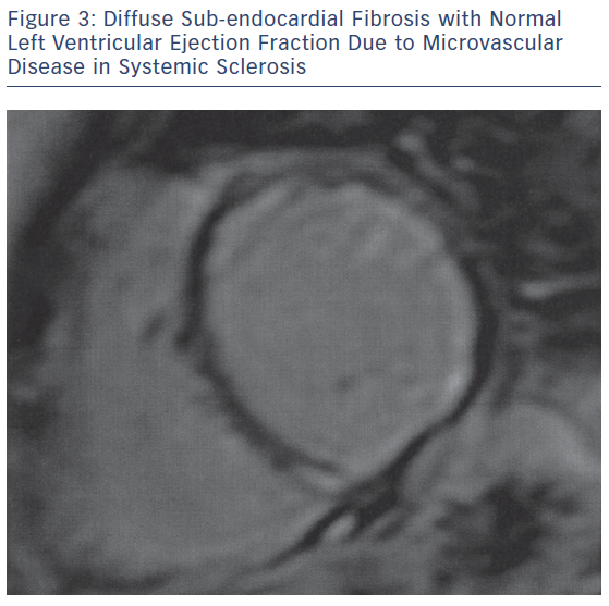
The main pathophysiologic phenomena that need to be assessed as early as possible during the course of ARDs include myocardial and vascular inflammation; macro- and micro-vascular vasculopathy; small epicardial, intra-myocardial and/or sub-endocardial fibrosis, due to inflammation and/or MI (Figures 1–3); acuity of heart involvement; angiography of the great vessels; and assessment of the arterial wall inflammatory process.11–15 These cannot be assessed by the currently used imaging modalities. It should be considered that most ARD patients are female and may be unable to exercise, due to arthritis or muscular discomfort or weakness and therefore the assessment of myocardial ischemia may be problematic.

The Need for a Cardio-rheumatology Outpatient Clinic

The multifaceted presentation of CVD in ARDs created the need for a specific outpatient cardio-rheumatic clinic. Our hospital was among the first in the world to create a dedicated clinic for early diagnosis, management and follow-up of CVD in ARDs. Our outpatient cardio-rheumatic clinic works in close collaboration with hospitals that diagnose and treat ARD patients.

Clinical examination, rest ECG, exercise ECG, echocardiography, nuclear techniques and cardiac catheterisation, if needed, are used as diagnostic tools. The exercise electrocardiogram is commonly used first line for diagnosing myocardial ischaemia. It is widely available and inexpensive, but has low sensitivity in women and is not diagnostic in patients with rhythm disturbances.16 The current American Heart Association/American College of Cardiology guidelines recommend the exercise electrocardiogram as the first-line test for known or suspected coronary disease, but only when the patient is able to exercise. However, ARD patients with musculoskeletal impairment are unable to exercise. The exercise electrocardiogram is also less accurate than alternative stress imaging technologies.17

Transthoracic echocardiography (TTE) is currently the cornerstone of diagnosing CVD. Stress echocardiography combines TTE with a physical, pharmacological or electrical stress, inducing higher pulse rate. If coronary stenosis is present, ischaemia of the left ventricular wall may occur, resulting in a transient worsening of left ventricular wall contractility. Stress echocardiography may provide similar diagnostic and prognostic accuracy with radionuclide stress perfusion imaging, but at a substantially lower cost, without environmental pollution, and with no bio-effects for the patient and the physician. However, it has the limitation of being an operator and acoustic window dependent technique.

Sub-epicardial Fibrosis in the Inferior Wall

Article image

Transmural MI of the Interventricular Septum

Article image

Stress myocardial perfusion scintigraphy (MPS) is a useful non-invasive imaging modality for diagnosing patients with suspected coronary artery disease.18,19 However, has serious limitations, including high radiation exposure, imaging artefacts and low spatial resolution that does not allow the detection of small areas of myocardial ischemia and scars.20 It became clear that all the above-mentioned modalities, although useful in cardiology, were unable to diagnose the multifaceted cardiac involvement in asymptomatic ARD patients.21

This discrepancy motivated the application of cardiovascular magnetic resonance (CMR). Compared with other non-invasive imaging modalities, CMR has the advantage of high spatial resolution capable of assessing slight tissue changes occurring during the course of ARDs. Additionally, CMR is versatile and can be used to perform functional, stress-rest perfusion, replacement and diffuse fibrosis assessment, all in one examination, without the use of ionising radiation.21 Furthermore, it can offer evaluation of the patency of the great, peripheral and coronary vessels. Recently, the use of mapping techniques and extracellular volume fraction has allowed the assessment of myocardial oedema and diffuse fibrosis in patients with renal impairment, which is commonly found in ARDs.21

Diffuse Sub-endocardial Fibrosis

Article image

The superiority of CMR in the diagnosis of CVD in ARDs against the other non-invasive imaging modalities is based on the assessment of:

  • myocardial ischaemia and/or sub-endocardial/transmural replacement or diffuse fibrosis, due to either macro- or micro-vascular coronary artery disease;22–26
  • disease acuity, due to macro- or micro-vascular coronary artery disease;22–27
  • extent and disease acuity of myocardial/vascular inflammation;27,28 and
  • aetiology of silent/overt heart failure or rhythm disturbances.29,30

The disadvantages of CMR are that it is contraindicated in patients with non-MRI compatible devices or metallic clips and in people with claustrophobia. However, all coronary artery stents, currently implanted valves and MRI-conditioned devices can be safely scanned. In patients with renal failure, the use of gadolinium is contraindicated, due to the risk of nephrogenic fibrosis. In these patients, we can apply non-contrast imaging protocols and perform both function and tissue characterisation evaluation. Additionally, CMR performs less well in patients with severe arrhythmia and who are unable to hold their breath.21

Role of CMR in the Evaluation of Anti-rheumatic and Cardiac Treatment

There are only a few studies supporting a role for CMR in the evaluation of anti-rheumatic and cardiac treatment in ARDs. A previous study by our group documented that CMR can detect early silent cardiovascular (CV) lesions, assess disease acuity and successfully evaluate the effect of both cardiac and anti-rheumatic medication on the CV system.31

In another study, the CMR findings of 246 ARD patients with typical cardiac symptoms (n=146) or atypical cardiac symptoms (n=100) were retrospectively evaluated. CMR in symptomatic ARD patients with normal echocardiographic findings assessed disease acuity and identified vasculitis, myocarditis and MI that influenced the CV risk stratification of ARD patients.11 Furthermore, occult CMR lesions, including myocardial oedema, myocarditis, diffuse sub-endocardial fibrosis and MI, were not unusual in treating naïve ARDs and may be reversed with appropriate treatment.32 Additionally, stress CMR has successfully detected silent myocardial Raynaud phenomena in patients with ARDs and known peripheral Raynaud phenomena, and thus motivated the early start of relevant cardiac treatment.33

CMR offers the potential to identify ARD patients at high risk of ventricular tachycardia or VF, thus influencing both cardiac and anti-rheumatic treatment and possibly modifying the criteria for ICD implantation.34 Although the role of cardiac treatment is established for early morphologic or functional cardiac changes,35 clear guidelines for anti-rheumatic treatment are still missing.

According to our experience, a baseline study, including clinical, ECG and TTE evaluation, should be performed at diagnosis of ARDs and a CMR should then be recommended if:

  • there is a mismatch between clinical findings and imaging/laboratory findings;
  • there is new onset heart failure;
  • if there is any kind of arrhythmia;
  • if the rheumatologist plans to change treatment, especially if there is a plan to initiate biologic agents;
  • if there is any increase in troponin, brain natriuretic peptide or D-dimers, even if there are only subtle symptoms;
  • if the patient is being treated with hydroxyl-chloroquine or biologic agents; or
  • if the patient notes any kind of typical or atypical cardiac symptoms and the routine cardiac evaluation is normal.

Conclusion

CMR allows the early diagnosis of various CV pathophysiologic phenomena in ARDs. Preliminary studies suggest it has a promising role in prompting modifications of anti-rheumatic and cardiac treatment in ARDs with CVD. The diagnostic potential of CMR strongly supports the view that we should “save the last dance” for CMR in early diagnosis, risk stratification and treatment follow-up of CVD in ARDs.

References

  1. Pohl D, Benseler S. Systemic inflammatory and autoimmune disorders. Handb Clin Neurol 2013;112:1243–52.
    Crossref | PubMed
  2. Aviña-Zubieta JA, Choi HK, Sadatsafavi M, et al. Risk of cardiovascular mortality in patients with rheumatoid arthritis: a meta-analysis of observational studies. Arthritis Rheum 2008;59:1690–7.
    Crossref | PubMed
  3. Sherer Y, Shoenfeld Y. Mechanisms of disease: atherosclerosis in autoimmune diseases. Nat Clin Pract Rheumatol 2006;2:99–106.
    Crossref | PubMed
  4. Kitas GD, Gabriel SE. Cardiovascular disease in rheumatoid arthritis: state of the art and future perspectives. Ann. Rheum Dis 2011;70:8–14.
    Crossref | PubMed
  5. Hollan I, Meroni PL, Ahearn JM, et al. Cardiovascular disease in autoimmune rheumatic diseases. Autoimmun Rev 2013;12:1004–15.
    Crossref | PubMed
  6. Björnådal L, Yin L, Granath F, et al. Cardiovascular disease a hazard despite improved prognosis in patients with systemic lupus erythematosus: results from a Swedish population based study 1964–95. J Rheumatol 2004;31:713–9.
    Crossref | PubMed
  7. Symmons DP, Gabriel SE. Epidemiology of CVD in rheumatic disease, with a focus on RA and SLE. Nat Rev Rheumatol 2011;7:399–408.
    Crossref | PubMed
  8. Gasparyan AY. Cardiovascular risk and inflammation: pathophysiological mechanisms, drug design, and targets. Curr Pharm Des 2012;18:1447–9.
    Crossref | PubMed
  9. Dimitroulas T, Giannakoulas G, Karvounis H, et al. Micro- and macrovascular treatment targets in scleroderma heart disease. Curr Pharm Des 2014;20:536–44.
    Crossref | PubMed
  10. Al-Dhaher FF, Pope JE, Ouimet JM. Determinants of morbidity and mortality of systemic sclerosis in Canada. Semin Arthritis Rheum 2010;39:269–77.
    Crossref | PubMed
  11. Mavrogeni S, Sfikakis PP, Gialafos E, et al. Cardiac tissue characterization and the diagnostic value of cardiovascular magnetic resonance in systemic connective tissue diseases. Arthritis Care Res (Hoboken) 2014;66:104–12.
    Crossref | PubMed
  12. Mavrogeni S, Spargias K, Markussis V, et al. Myocardial inflammation in autoimmune diseases: investigation by cardiovascular magnetic resonance and endomyocardial biopsy. Inflamm Allergy Drug Targets 2009;8:3907.
    Crossref | PubMed
  13. Mavrogeni S, Manoussakis MN. Myocarditis and subclavian stenosis in Takayasu arteritis. Int J Cardiol 2011;148:223–4.
    Crossref | PubMed
  14. Mavrogeni S, Sfikakis PP, Gialafos E, et al. Diffuse, subendocardial vasculitis. A new entity identified by cardiovascular magnetic resonance and its clinical implications. Int J Cardiol 2013;168:2971–2.
    Crossref | PubMed
  15. Raman SV, Aneja A, Jarjour WN. CMR in inflammatory vasculitis. J Cardiovasc Magn Reson 2012;14:82.
    Crossref | PubMed
  16. Meyers HP, Jaffa E, Smith SW, et al. Evaluation of T-wave morphology in patients with left bundle branch block and suspected acute coronary syndrome. J Emerg Med 2016;51:229-37.
    Crossref | PubMed
  17. Gibbons RJ, Balady GJ, Beasley JW et al. ACC/AHA guidelines for exercise testing. J Am Coll Cardiol 1997;30:260–311. PMID: 9207652.
    Crossref | PubMed
  18. Sicari R, Nihoyannopoulos P, Evangelista A, et al. European Association of Echocardiography. Stress echocardiography expert consensus statement: European Association of Echocardiography (EAE). Eur J Echocardiogr 2008;9:415–37.
    Crossref | PubMed
  19. Hachamovitch R, Berman DS, Kiat H, et al. Exercise myocardial perfusion SPECT in patients without known coronary artery disease. Circulation 1996;93:905–14.
    PubMed
  20. Iskander S, Iskandrian AE. Risk assessment using single-photon emission computed tomography technetium-99m sestamibi imaging. J Am Coll Cardiol 1998;32:57–62.
    Crossref | PubMed
  21. Mavrogeni SI, Kitas GD, Dimitroulas T, et al. Cardiovascular magnetic resonance in rheumatology: Current status and recommendations for use. Int J Cardiol 2016;217:13548.
    Crossref | PubMed
  22. Mavrogeni S, Sfikakis PP, Gialafos E, et al. Cardiac tissue characterization and the diagnostic value of cardiovascular magnetic resonance in systemic connective tissue diseases. Arthritis Care Res (Hoboken) 2014;66:10412.
    Crossref | PubMed
  23. Mavrogeni S, Markousis-Mavrogenis G, Koutsogeorgopoulou L, et al. Cardiovascular magnetic resonance imaging pattern at the time of diagnosis of treatment naïve patients with connective tissue diseases. Int J Cardiol 2017;236:1516.
    Crossref | PubMed
  24. Chraibi S, Ibnabdeljalil H, Habbal R, et al. Pericardial tamponade as the first manifestation of dermatopolymyositis. Ann Med Interne (Paris) 1998;149:4646.
    PubMed
  25. Mavrogeni S, Bratis K, Sfendouraki E, et al. Myopericarditis, as the first sign of rheumatoid arthritis relapse, evaluated by cardiac magnetic resonance. Inflamm Allergy Drug Targets 2013;12:20611.
    Crossref | PubMed
  26. Mavrogeni SI, Schwitter J, Gargani L, et al. Cardiovascular magnetic resonance in systemic sclerosis: “Pearls and pitfalls”. Semin Arthritis Rheum 2017;47:7985.
    Crossref | PubMed
  27. Kouranos V, Tzelepis GE, Rapti A, et al. Complementary role of CMR to conventional screening in the diagnosis and prognosis of cardiac sarcoidosis. JACC Cardiovasc Imaging 2017;10:143747.
    Crossref | PubMed
  28. Raman SV, Aneja A, Jarjour WN. CMR in inflammatory vasculitis. J Cardiovasc Magn Reson 2012;14:82.
    Crossref | PubMed
  29. Mavrogeni S, Sfikakis PP, Karabela G, et al. Cardiovascular magnetic resonance imaging in asymptomatic patients with connective tissue disease and recent onset left bundle branch block. Int J Cardiol 2014;171:827.
    Crossref | PubMed
  30. Kobayashi Y, Kobayashi H, Giles JT, et al. Association of tocilizumab treatment with changes in measures of regional left ventricular function in rheumatoid arthritis, as assessed by cardiac magnetic resonance imaging. Int J Rheum Dis 2016;19:116974.
    Crossref | PubMed
  31. Mavrogeni S, Markousis-Mavrogenis G, Koutsogeorgopoulou L, Kolovou G. Cardiovascular magnetic resonance imaging: clinical implications in the evaluation of connective tissue diseases. J Inflamm Res 2017;10:5561.
    Crossref | PubMed
  32. Mavrogeni S, Markousis-Mavrogenis G, Koutsogeorgopoulou L, et al. Cardiovascular magnetic resonance imaging pattern at the time of diagnosis of treatment naïve patients with connective tissue diseases. Int J Cardiol 2017;236:1516.
    Crossref | PubMed
  33. Mavrogeni S, Bratis K, Koutsogeorgopoulou L, et al. Myocardial perfusion in peripheral Raynaud’s phenomenon. Evaluation using stress cardiovascular magnetic resonance. Int J Cardiol 2017;228:4448.
    Crossref | PubMed
  34. Mavrogeni SI, Sfikakis PP, Dimitroulas T, et al. Prospects of using cardiovascular magnetic resonance in the identification of arrhythmogenic substrate in autoimmune rheumatic diseases. Rheumatol Int 2018;38:161521.
    Crossref | PubMed
  35. Ponikowski P, Voors AA, Anker SD, et al. 2016 ESC guidelines for the diagnosis and treatment of acute and chronic heart failure. Eur J Heart Fail 2016;18:891975.
    Crossref | PubMed